


Introduction
Root resorption is a gradual loss of the dentin and cementum, inevitably leading to its atrophy [1]. The roots of teeth then become shorter and thinner. The fibres that hold the tooth in the bone are also damaged, which causes a significant increase in mobility and results in possible tooth loss. While in the case of deciduous teeth, root resorption is an appropriate situation that allows the child to replace the milk teeth with permanent ones, in adults it is considered a pathological process. Based on the location, tooth resorption is divided into internal, external, and external-internal. External resorption can be classified into 5 different types [2,3]. One of them is external inflammatory apical root resorption, which can be caused by orthodontic treatment. In 1988 Levander and Malmgren proposed a classification considering the degree of root length loss (Table 1) [4].
Table 1
Levander and Malmgren classification of root resorption
The risk of orthodontically induced external apical root resorption (OIEARR) is significant, and it depends on many aspects like type of malocclusion, treatment method, duration of the treatment, or systemic diseases [5-7]. Dentists and especially orthodontists should be aware of the danger and monitor the length of the tooth roots in order to respond appropriately or stop the treatment when necessary. A study performed by Remigton et al. [8] showed in long-term evaluation that resorption does not progress significantly after the end of treatment. Root resorption is usually detected accidentally during routine radiological examination.
The aim of this investigation was to evaluate the frequency of apical root resorption in the anterior teeth of the maxilla visible on panoramic images during ortho-dontic treatment with a fixed appliance according to Levander and Malmgren classification.
Material and methods
A total of 194 panoramic radiographs of patients with a fixed appliance in the upper arch were obtained from the database of the Department of Dental and Maxillofacial Radiodiagnostics of the Medical University of Lublin. Then, all the images were analysed by 2 independent observers to evaluate the occurrence and severity of root resorption in maxillary incisors and canines during ortho-dontic treatment, in its final stages. All examinations were taken with a VistaVoxS Panoramic Unit (Dürr Dental, Germany). Age, gender, and the number and name of teeth with external root resorption were collected. The research group included 135 females and 59 males, aged 15-28 years with a mean 20.6 years. The degree of root resorption was assessed according to the Levander and Malmgren classification.
Results
Considering all examined panoramic images, there were 146 patients with variable stages of root resorption (75.26%). In the group of females, we found that 103 patients had external resorption (76.29%) that was slightly higher than the findings in the group of men (72.88%). Age was not found to be a significant factor in our study.
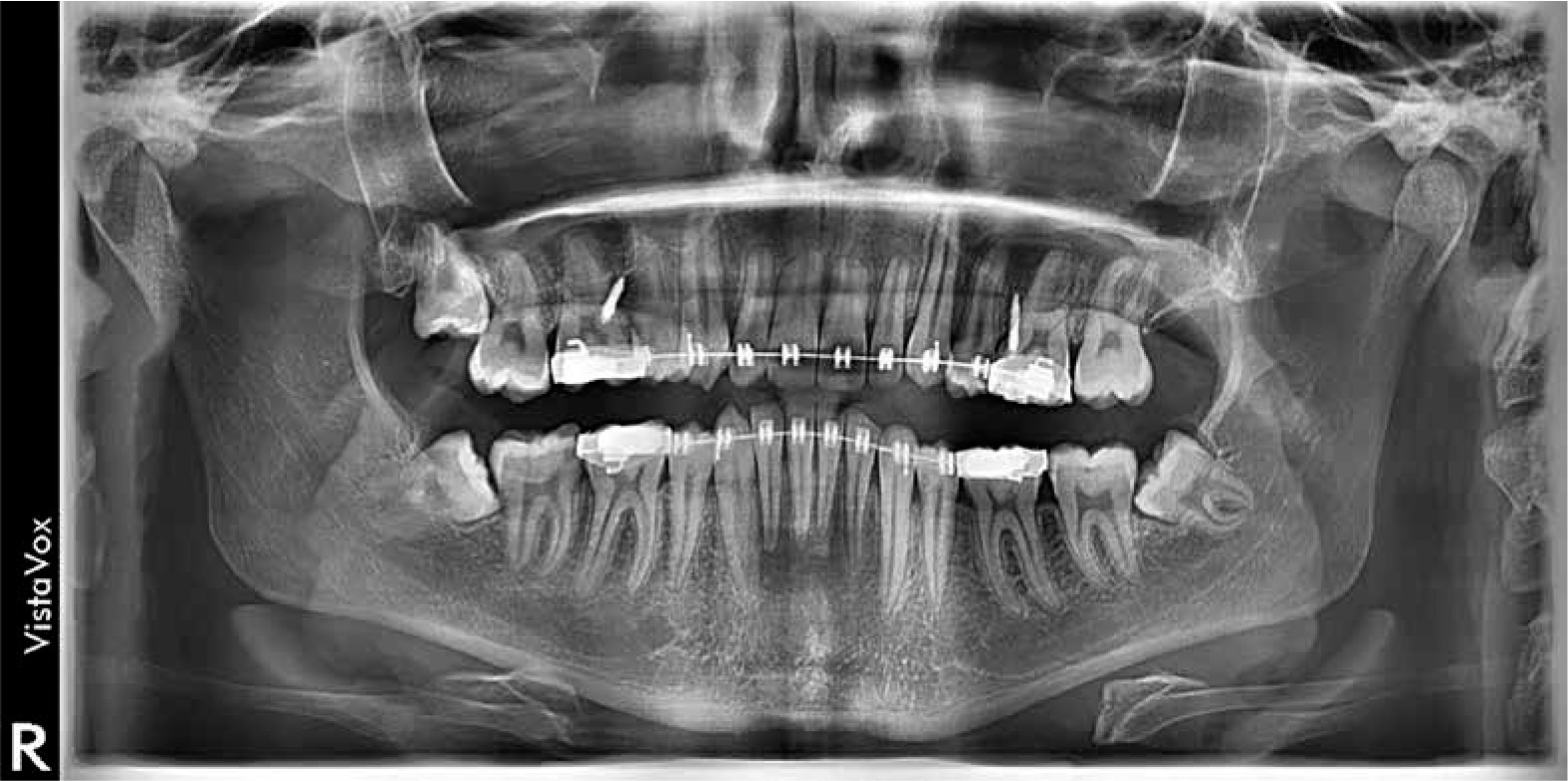
Among 1164 evaluated anterior teeth, 469 showed signs of resorption (40.3%). The number of teeth with different degrees of root destruction is shown in Table 2. The tooth most frequently affected by resorptive changes was the right central upper incisor [11]. Figure 1 shows a panoramic radiograph with visible resorption of maxillary incisors (III and IV stage). The most common stage of root resorption was stage II (53.09%) according to Levander classification. The fourth stage was the least common (2.99%), and 78.57% of the teeth with this stage were central incisors.
Discussion
Root resorption can be diagnosed on periapical radiographs [9,10], panoramic images [11,12], or cone beam computed tomography (CBCT) [13]. The most common radiological examinations use in diagnosing and planning in orthodontics are panoramic radiographs and cephalograms [14]. Panoramic radiograph has a lot of advantages, including visibility of the entire dental arch and lower doses of radiation in comparison with CBCT. However, it also has some limitations, like overlapping different anatomical structures or magnification of the image. A study comparing panoramic and periapical radiographs in detecting external root resorption showed that on panoramic radiographs the stage of root resorption is significantly higher [10]. The authors suggested that it might be caused by magnification of the image, which is 20-35% on average. A similar study concluded that periapical images are more efficient in the assessment of the root shape and level of resorption [15]. CBCT seems to be the most precise radiological examination due to the possibility of 3-dimensional evaluation. There are many studies that show predominance of CBCT over different radiological tools in root resorption assessment [16,17]. However, following the rule ALARA (as low as reasonably achievable) it is necessary to avoid an additional radiation exposure, and panoramic images seem to be sufficient for detecting and diagnosing external root resorption.
Janson et al. [18] presented a study that evaluated apical resorption by use of periapical radiographs after treatment with 3 different fixed appliance techniques; the results showed that in the whole sample the most frequently affected teeth were upper central incisors, which coincides with our research, but it is not with the agreement with previous studies that indicated that lateral incisors have a greater predisposition to resorptive changes [19-21]. Jiang et al. [22], in a study performed using panoramic radiographs, found that central incisors were the primarily resorbed teeth, which is compatible with our results. According to Elhaddaoui et al. [23], root resorption after orthodontic treatment measured on panoramic radiograph is usually lower than 2.5 mm with less than a 20% chance of severe resorption. In our study the most common type of external root resorption was a second degree according to Levander and Malmgren. There were 361 teeth with stage I or II RR, which means that 77% of examined teeth had a root destruction up to 2 mm. This is in agreement with the study of Elhaddaoui [23]. The Authors claim that severe root resorption mainly concerns maxillary lateral incisors, which contrasts with our finding that 78.57% of stage IV (with resorption above 1/3 of root) was detected in upper central incisors. Overall, both lateral and central maxillary incisors are the most susceptible to resorption, probably due to the shape of the root (bottle shaped or blunted) [4,24].
A study based on CBCT measurements [25] showed that 6.6% of patients had at least one tooth with extreme resorption (above 4 mm). The systematic review performed by Weltman [26] showed that severe orthodontically induced external root resorption appeared in only 1-5% of the teeth, which agrees with our findings (2.99%). Previous studies showed that prevalence of root resorption greater than 2 mm (stage III and IV) varied from 10% to 18% [4,27]. In another study, Levander and Malmgren [28] examined patients with oligodontia and found that only 5% of teeth were resorbed in 2 mm and more. The authors suggested that it might be caused by a high proportion of missing upper lateral incisors in the sample.
A study from Poland Kowalska et al. [29] measured the width-to-length ratio in incisor roots visible in panoramic view before and after orthodontic treatment. The authors evaluated upper and lower incisors, and they showed that only 10.83% of the examined teeth presented with signs of resorption. This low value could be caused by a reduction of orthodontic forces during the treatment in comparison to older techniques using light wires.
When gender is taken into account, there are many studies that showed no significant correlation of sex and the prevalence or amount of external resorption with orthodontic treatment [19,21,23,25,28,30-32], which is similar to our findings. However, in a study from 1975 Newman indicated that females are more susceptible to apical root loss than males [33]. According to some previous studies [22,27] patient’s age may be a factor influencing the prevalence and level of resorptive changes in maxillary anterior teeth, which is contrary to the results of Sameshima and Sinclair [31,34] who found that adults experienced more advanced resorption than children but only in the mandibular anterior teeth. The authors did not find correlation between age and the amount of apical root resorption in the maxillary anterior region. They also showed that the most affected teeth were lateral upper incisors and those with abnormal shape of the root. Additionally, they found that the Caucasian population is more prone to experience greater root resorption than Asian patients.
Panoramic radiographs can be used in diagnosing and detecting EARR, but further studies should be performed including 3D imaging (CBCT), which seems to be more accurate in the evaluation of small structures and details.
Conclusions
External apical root resorption due to orthodontic treatment is a common finding on panoramic radiographs. In our study, maxillary central incisors were the most frequently affected teeth, followed by lateral upper incisors and canines. The gender and age of the patients were not found to be significant factors increasing the risk of root resorption.