ISSN: 1899-0967
Polish Journal of Radiology
Established by prof. Zygmunt Grudziński in 1926 Sun
Current issue Archive Manuscripts accepted About the journal Editorial board Abstracting and indexing Contact Instructions for authors Ethical standards and procedures
Editorial System
Submit your Manuscript
SCImago Journal & Country Rank
1/2019
vol. 84
 
Share:
Share:
Chest radiology
Original paper

The diagnostic accuracy of endobronchial ultrasound and spiral chest computed tomography scan in the prediction of infiltrating and non-infiltrating lymph nodes in patients undergoing endobronchial ultrasound

Sepideh Haghi
1
,
Shahram Kahkouee
2
,
Arda Kiani
3
,
Atefeh Abedini
3
,
Neda Akhoundi
1
,
Mehdi Javanbakht
4
,
Hadi Rezaei
1
,
Mersede Paraham
1

1.
College of Medicine, Shahid Beheshti University of Medical Sciences, Iran
2.
Department of Radiology, Masih Daneshvari Hospital, Shahid Beheshti University of Medical Sciences, Iran
3.
Department of Pulmonary Medicine, Medical University of Shahid Beheshti, Iran
4.
Economics Unit, Device Access UK Ltd., University of Southampton Science Park, Chilworth, Southampton, United Kingdom
© Pol J Radiol 2019; 84: e565-e569
Online publish date: 2019/12/19
Article file
Get citation
 
PlumX metrics:
 

Introduction

Mediastinal and hilar lymphadenopathy is usually diagnosed by computerised tomography (CT) scan, which is also used for staging, with accuracy and specificity of 57% and 82%, respectively [1]. In surgical staging approximately 18% of patients with a negative CT scan develop metastatic mediastinal disease [2]. However, all lymph nodes may not be detected. In these cases, usually thoracoscopy and anterior mediastinoscopy are used [2]. But all these methods for staging are mainly invasive and would result in high costs [3,4]. However, the positron emission tomography (PET) scan may develop high applicability for staging of metastatic lung cancer with lymph node involvement [5], but it is less frequently used due to high costs [6]. Endoscopic ultrasound-guided fine needle aspiration (FNA) was also introduced; the main limitation for this method is the impossibility for airway assessment [7-10].

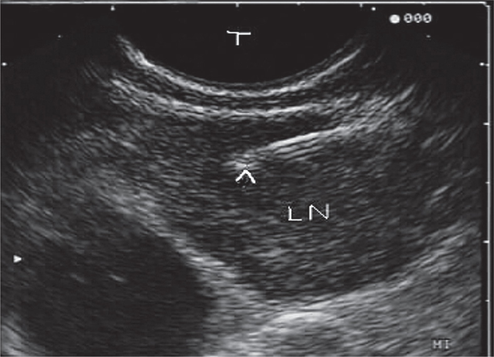
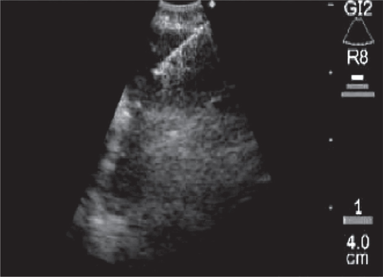
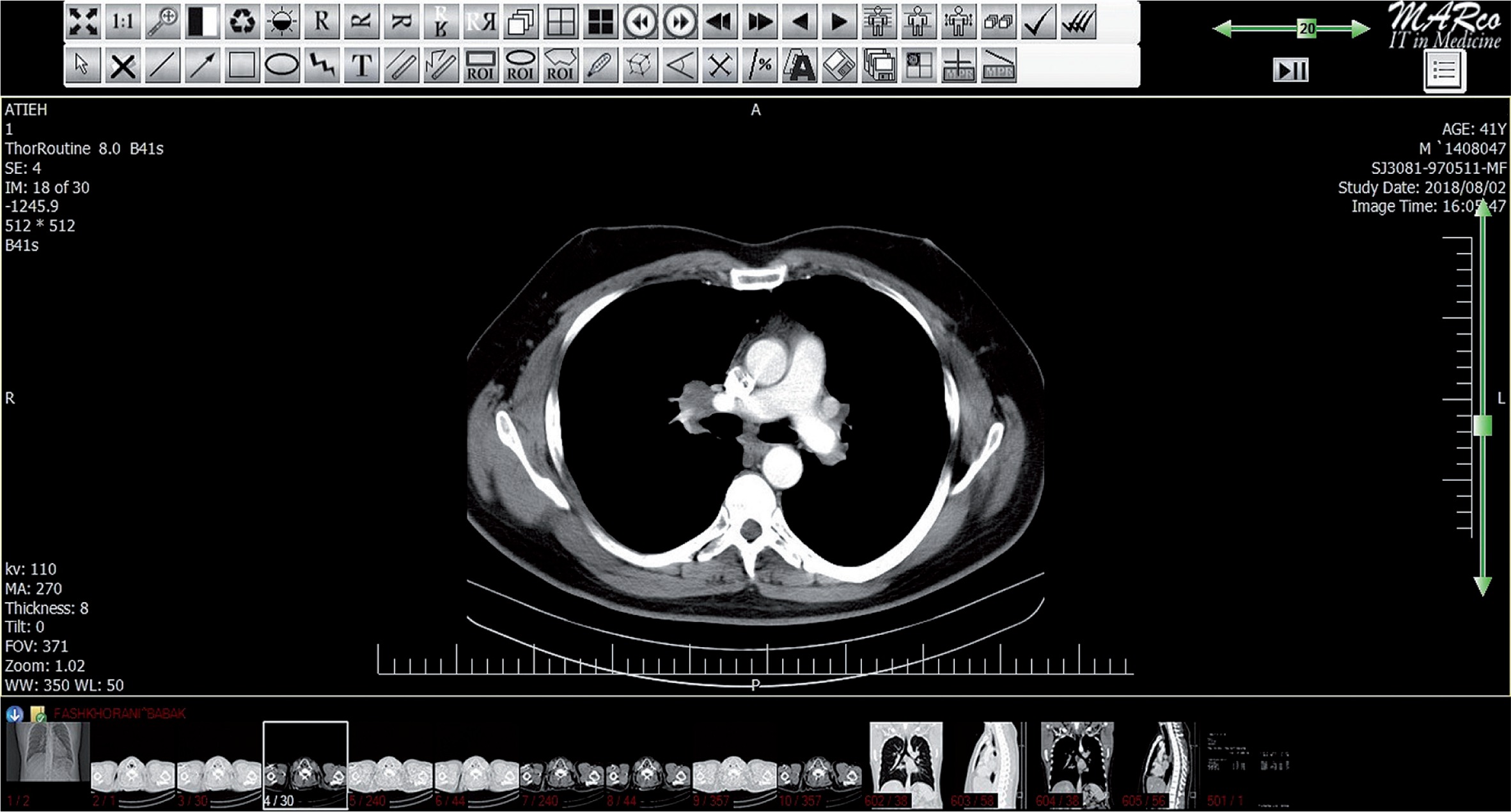
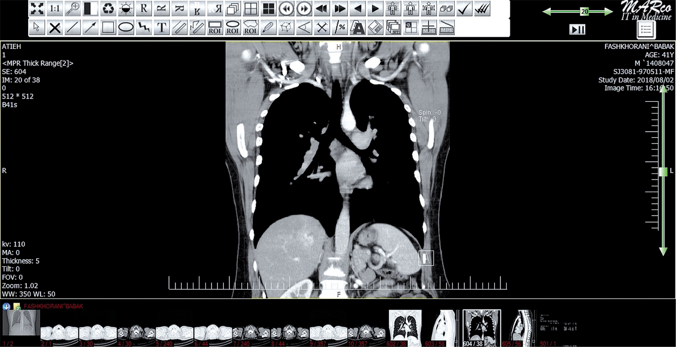
In this era some studies have reported highest applicability in cases using endobronchial ultrasonography (EBUS) with high accuracy in false negative CT scan [11-14]. However, there are some controversies about the role of the EBUS (Figures 1 and 2) versus spiral chest CT scan (Figures 3 and 4) to differentiate between infiltrated and none-infiltrated lymph nodes.

Figure 1

Endobronchial ultrasound fine needle aspiration paratracheal lymph node

/f/fulltexts/PJR/39489/PJR-84-39489-g001_min.jpg
Figure 2

Endobronchial ultrasound fine needle aspiration hilar lymph node

/f/fulltexts/PJR/39489/PJR-84-39489-g002_min.jpg
Figure 3

Hilar lymph nodes in axial computed tomography

/f/fulltexts/PJR/39489/PJR-84-39489-g003_min.jpg
Figure 4

Hilar and subcarinal lymph nodes in coronal computed tomography

/f/fulltexts/PJR/39489/PJR-84-39489-g004_min.jpg

Hence, for improvement in diagnostic approaches, the purpose in this study was to determine the diagnostic accuracy of endobronchial ultrasound and spiral chest CT scan in the prediction of infiltrating and non-infiltrating lymph nodes in patients undergoing endobronchial ultrasound.

Material and methods

In this observational prospective study, 40 consecutive patients with infiltrating and non-infiltrating lymph nodes in Masih-Daneshvari Hospital, Tehran, Iran in 2017 and 2018, who had positive pathology results by biopsy or EBUS but with non-diagnostic spiral chest CT scan, were enrolled. The exclusion criterion was incomplete data.

The study was approved by the local Ethical Committee. Data were collected by existing medical documents and checklist among understudy patients. Also, the pathological findings including benign and malignant status were recorded in the patients. The size and location of the lymph nodes were also determined.

Data analysis was done with SPSS software (Version 25.0), and mean plus standard deviation were calculated for numerical variables, and the frequency and percentage were reported for categorical factors. Kappa test was used to determine the congruence, and the sensitivity, specificity, and accuracy was calculated. P values less than 0.05 were considered statistically significant.

Results

The patient’s age was under 50 years in 17 cases (42.5%). The gender was male in 15 patients (37.5%). The CT scan results were positive in 33 patients (82.5%), and the EBUS was positive in 35 cases (87.5%). According to the pathological results, 31 cases (77.5%) were benign. The mean lymph node size was 10.7 mm in CT and 11 mm in EBUS without difference between benign or malignant lesions (p > 0.05). Also, the total range was from 6 to 25 mm.

The results in this study (Table 1) show that the congruence between EBUS and CT scan was 80.5% (p = 0.0001). The sensitivity, specificity, and accuracy for CT scan was 100%, 22.6%, and 40%, respectively (Table 2) and the sensitivity, specificity, and accuracy for EBUS was 100%, 16.1%, and 35%, respectively (Table 3).

Table 1

Congruence between computed tomography scan and endobronchial ultrasound results

CTTotal
PosNeg
EBUSPosCount33235
 % within EBUS94.3%5.7%100%
 % within CT100%28.6%87.5%
NegCount055
 % within EBUS0100%100%
 % within CT071.4%12.5%
TotalCount33740
 % within EBUS82.5%17.5%100%
 % within CT100%100%100%
Table 2

Computed tomography scan sensitivity, specificity, and accuracy

Pathological resultTotal
BenignMalignant
EBUSPosCount24933
 % within CT72.7%27.3%100%
 % within77.4%100%82.5%
 Pathological result   
NegCount707
 % within CT100%0100%
 % within22.6%017.5%
 Pathological result   
TotalCount31940
 % within CT77.5%22.5%100%
 % within100%100%100%
 Pathological result   
Table 3

Endobronchial ultrasound sensitivity, specificity, and accuracy

Pathological resultTotal
BenignMalignant
EBUSPosCount26935
 % within EBUS74.3%27.5%100%
 % within83.9%100%87.5%
 Pathological result   
NegCount505
 % within EBUS100%0100%
 % within16.1%012.5%
 Pathological result   
TotalCount31940
 % within EBUS77.5%22.5%100%
 % within100%100%100%
 Pathological result   

The congruence did not differ according to age, and patients younger and older than 50 years had good congruence between EBUS and CT scan. Also, the congruence did not differ according to gender, and both male and female patients had good congruence between EBUS and CT scan. The congruence was good in benign cases (p = 0.0001), but it was not determined according to the location, malignancy, and stage of the malignancy (p > 0.05).

Discussion

The main utilised method to diagnose mediastinal lymphadenopathy is CT scan, but it would show low accuracy. The highest applicability point is related to EBUS. However, there are some controversies. Because CT scan is a non-invasive method, in this study the accuracy of EBUS and spiral CT scan was assessed. The congruence of methods was 80.5% and significant. The sensitivity for methods was 100% and the specificity was near to 20% with accuracy rate of 35 to 40%. Age and gender subgroups both had good equal concordances. The congruence was higher in benign lesions. It is not clear in malignant lesions. Also, the location and stage effects were impossible to determine. Udoji et al. [15] reported that lymph nodes had larger size in EBUS versus CT scan assessment. In our study, despite a lack of difference between benign and malignant lesions, it was larger in EBUS evaluation.

Helmy et al. [16] assessed 11 patients who were suspected of having lymph node malignancy without CT scan positive results. The malignancy was reported in 54.4% of cases under bronchoscopy, 18.2% under EBUS-TBNA, and 27.3% under CT-guided biopsy. From 11 cases with negative CT scan the EBUS was positive in four patients [16]. In our study the malignancy rate was lower, but similarly EBUS had good applicability.

Nakajima et al. [17] showed that among 106 patients with metastatic lung cancer the sensitivity and specificity were 92% and 100%, respectively. Their sensitivity and specificity were lower and higher than ours, respectively. Memoli et al. [18] reported malignancy in lymph nodes in 22.5% of cases and reported good diagnostic applicability for both CT scan and EBUS method as well as our study.

Jhun et al. [19] reported sensitivity and specificity of 92% and 99%, respectively, for EBUS, which was related to lymph node location. However, in our study, due to the small number of hilar samples, a comparison was impossible. Lee et al. [20] assessed 247 patients with hilar and mediastinal lymphadenopathy and reported that older age and pathological results were related to higher false negative rates. In our study the benign status had some effects on congruence between methods, but there was no difference for age.

According to the obtained results it may be concluded that CT scan and EBUS results have good congruence and high sensitivity to differentiate infiltrating and non-infiltrating lymph nodes. Hence, these methods are useful for screening methods, but due to their low specificity and accuracy their use for a confirmative approach is not beneficial. However, regarding its accessibility and less invasive nature, the use of chest CT scan is more rational and is recommended in these patients. However further studies with larger sample sizes and controlling for confounding factors would develop more definite results.

Disclosure

The authors declare no conflicts of interest.

References

1 

Toloza EM, Harpole L, Detterbeck F, McCrory DC , authors. Invasive staging of non-small cell lung cancer: a review of the current evidence. Chest. 2003. 123:p. 157–166

2 

Hoffmann H , author. Invasive staging of lung cancer by mediastinoscopy and video-assisted thoracoscopy. Lung Cancer. 2001. 34:p. 3–5

3 

Luke WP, Pearson FG, Todd TR, et al. , authors. Prospective evaluation of mediastinoscopy for assessment of carcinoma of the lung. J Thorac Cardiovasc Surg. 1986. 91:p. 53–56

4 

Coughlin M, Deslauriers J, Beaulieu M, et al. , authors. Role of mediastinoscopy in pretreatment staging of patients with primary lung cancer. Ann Thorac Surg. 1985. 40:p. 556–560

5 

Toloza EM, Harpole L, McCrory DC , authors. Noninvasive staging of non-small cell lung cancer: a review of the current evidence. Chest. 2003. 123 Suppl 1:p. 137–146

6 

Brim O, Kappetein AP, Stijnen T, Bogers AJ , authors. Meta-analysis of positron emission tomographic and computed tomographic imaging in detecting mediastinal lymph node metastases in nonsmall cell lung cancer. Ann Thorac Surg. 2005. 79:p. 375–382

7 

Jacobson BC, Hirota WK, Goldstein JL, et al. , authors. The role of EUS for evaluation of mediastinal adenopathy. Gastrointest Endoscopy. 2003. 58:p. 819–821

8 

Wallace MB, Woodward TA, Raimondo M , authors. Endoscopic ultrasound and staging of non-small cell lung cancer. Gastrointest Endosc Clin N Am. 2005. 5:p. 157–167

9 

Annema JT, Versteegh MI, Veselic M, et al. , authors. Endoscopic ultrasound-guided fine-needle aspiration in the diagnosis and staging of lung cancer and its impact on surgical staging. J Clin Oncol. 2005. 23:p. 8357–8361

10 

Annema JT, Versteegh MI, Veselic M, et al. , authors. Endoscopic ultrasound added to mediastinoscopy for preoperative staging of patients with lung cancer. JAMA. 2005. 294:p. 931–936

11 

Krasnik M, Vilmann P, Larsen SS, Jacobsen GK , authors. Preliminary experience with a new method of endoscopic transbronchial real time ultrasound guided biopsy for diagnosis of mediastinal and hilar lesions. Thorax. 2003. 58:p. 1083–1088

12 

Yasufuku K, Chiyo M, Sekine Y, et al. , authors. Real-time endobronchial ultrasound-guided transbronchial needle aspiration of mediastinal and hilar lymph nodes. Chest. 2004. 126:p. 122–128

13 

Rintoul RC, Skwarski KM, Murchison JT, et al. , authors. Endobronchial and endoscopic ultrasound-guided real-time fine-needle aspiration for mediastinal staging. Eur Respir J. 2005. 25:p. 416–421

14 

Okamoto H, Watanabe K, Nagatomo A, et al. , authors. Endobronchial ultrasonography for mediastinal and hilar lymph node metastases of lung cancer. Chest. 2002. 121:p. 1498–1506

15 

Udoji TN, Phillips GS, Berkowitz EA, et al. , authors. Mediastinal and hilar lymph node measurements. comparison of multidetector-row computed tomography and endobronchial ultrasound. Ann Am Thorac Soc. 2015. 12:p. 914–920

16 

Helmy NA, Kamel KM, Naglaa BA , authors. Endobronchial ultrasonography for lung cancer staging in negative mediastinum on computed tomography. Egypt J Chest Dis Tuberc. 2013. 62:p. 475–479

17 

Nakajima T, Yasufuku K, Iyoda A, et al. , authors. The evaluation of lymph node metastasis by endobronchial ultrasound-guided transbronchial needle aspiration: crucial for selection of surgical candidates with metastatic lung tumors. J Thorac Cardiovasc Surg. 2007. 134:p. 1485–1490

18 

Wang Memoli JS, El-Bayoumi E, Pastis NJ, et al. , authors. Using endobronchial ultrasound features to predict lymph node metastasis in patients with lung cancer. Chest. 2011. 140:p. 1550–1556

19 

Jhun BW, Park HY, Jeon K, et al. , authors. Nodal stations and diagnostic performances of endobronchial ultrasound-guided transbronchial needle aspiration in patients with non-small cell lung cancer. J Korean Med Sci. 2012. 27:p. 46–51

20 

Lee J, Kim YK, Seo YY, et al. , authors. Clinical characteristics of false-positive lymph node on chest CT or PET-CT confirmed by endobronchial ultrasound-guided transbronchial needle aspiration in lung cancer. Tuberc Respir Dis (Seoul). 2018. 81:p. 339–346

Copyright: © Polish Medical Society of Radiology This is an Open Access article distributed under the terms of the Creative Commons Attribution-Noncommercial-No Derivatives 4.0 International (CC BY-NC-ND 4.0). License allowing third parties to download articles and share them with others as long as they credit the authors and the publisher, but without permission to change them in any way or use them commercially.
 
Quick links
© 2024 Termedia Sp. z o.o.
Developed by Bentus.