Introduction
Uterine artery embolization (UAE) is a safe, effective, and minimally invasive method for the treatment of uterine leiomyomas and their vast symptomatology, including ade-nomyosis [1,2]. Uterine leiomyomas, the most common benign tumour of premenopausal women, can cause extensive symptoms such as menorrhagia, dysmenorrhoea, dyspareunia, urinary frequency, and urinary urgency [2]. Although traditionally managed by total abdominal or laparoscopic hysterectomy, the advent of UAE has allowed for a minimally invasive treatment modality that also spares the uterus [2,3]. UAE not only preserves fertility but also minimizes patient recovery time and the rate of surgical complications. Multiple prior studies have established UAE to be effective in controlling haemorrhage and mitigating bulk-related symptoms [4-6]. For example, the FIBROID registry, a prospective study encompassing 72 practices varying in size and experience, demonstrated low 30-day complication rates, and a statistically significant improvement in reported symptoms and quality of life at 36 months [7,8].
Initially introduced in 1974, conventional UAE has been performed via transfemoral arterial access (TFA) [9]. Over the years, transradial arterial access (TRA) has gained traction in body interventional literature and has shown numerous advantages over TFA. Many of these studies have touted TRA to be superior to TFA due to reduced equipment costs, shorter lengths of post-procedural hospital stay, and decreased access site complications [10-13]. Additionally, TRA has been shown as the preferred route of access for patients in regards to overall comfort, pain levels, and early ambulation [14,15]. Although limited, prior literature has established TRA as a feasible approach to perform UAE, reporting a 100% technical success rate [16]. Despite this, TRA has been associated with a small yet significant increase in radiation exposure in both diagnostic and interventional procedures, potentially contributing to the general hesitance seen in adopting it as an option among operators [10-13].
To date, there have been limited studies in the body of interventional literature comparing transradial and transfemoral access in regard to pertinent radiation parameters: peak radiation dose, fluoroscopy time, procedure time, contrast volume, and equipment cost. The purpose of this study is to evaluate the potential benefits and pitfalls between the 2 different vascular approaches (TRA vs. TFA) in the specific population of patients who have undergone UAE for the treatment of fibroids.
Material and methods
This retrospective study, which was performed under clinical study guidelines, was approved by our institutional review board and was found to be IRB exempt. The demographic information and radiation-related data were collected based on a combination of electronic medical records and radiation safety worksheets for each IR procedure. A total of 172 patients underwent UAE procedures at our institute between October 2014 and June 2020. These patients were retrospectively reviewed within 2 groups: transradial artery approach (96 patients, mean age 43.2 ± 7.18 years, median age 44 years, total 96 procedures) and transfemoral artery approach (76 patients, mean age 43.7 ± 7.23 years, median age 44 years, total 76 procedures). The choice of procedure was based on the preferences of 7 different operators with experience ranging from 2 to more than 20 years serving in the interventional radiology faculty at a tertiary core hospital. These 7 operators conducted equal amounts of TRA and TFA procedures as each other throughout our study, proving no inconsistencies in training with respect to each subset. They also all began learning TRA in 2014 with its introduction at our institution. All procedures for both TRA and TFA were conducted in the same up-to-date angiography system (Siemens Healthineers, Malvern, PA).
Transradial artery approach
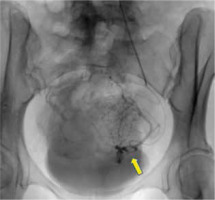
Typical TRA UAE was performed after Barbeau’s eva-luation of the radial artery [17]. Patients with a type D response were excluded from the study. For every patient, an ultrasound image documented the radial artery to be 2 mm in size. Prior to the procedure, the skin overlying the left radial artery was anaesthetized with lidocaine and nitroglycerin paste. Under ultrasound guidance, the radial artery was accessed with a 21-gauge needle. After placement of a 5F vascular access sheath, a 5F angled tip hydrophilic Glidecath (Terumo, Tokyo, Japan) was advanced to the internal iliac artery. Through this, a Renegade Hi-Flo microcatheter was advanced (Boston Scientific, Natick, MA) and used to select the uterine artery (Figure 1). For each patient, a radial artery “cocktail” was utilized post-procedure which included 200 ug nitroglycerin, 2.5 mg verapamil, and 3000 units of heparin.
Figure 1
Transradial access for uterine artery embolization. Typical case using a 5-F, angled-tip, hydrophilic-coated Glidecath (Terumo, Tokyo, Japan) and Renegade Hi-Flo (Boston Scientific, Natick, MA) microcatheter to access the horizontal segment of the uterine artery (yellow arrow)

After embolization, all wires and catheters were removed. Before removal of the sheath, a TR Band (Terumo, Somerset, NJ) was placed on the left wrist over the arteriotomy site and inflated to obtain haemostasis. The haemostasis was subsequently maintained for 60 minutes. Arterial haemostasis was reconfirmed as the cuff was incrementally deflated. Upon cuff removal by nursing staff in the recovery unit, the patient was observed for an additional 30 minutes prior to discharge.
Transfemoral artery approach
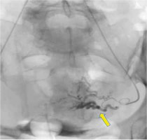
Under ultrasound guidance, typical TFA UAE was performed by placement of a 5F vascular access sheath, through which a 5F RIM catheter (Angiodynamics, Latham, NY) was advanced to the contralateral internal iliac artery. Through this, a 3F Renegade Hi-Flo microcatheter was advanced (Boston Scientific, Natick, MA) and used to select the uterine artery (Figure 2). Embolization was performed using 500-700 micron particles to stasis.
Figure 2
Transfemoral access for uterine artery embolization. Typical case will use a 5-F hydrophilic-coated RIM catheter (Angiodynamics, Latham, NY) and Renegade Hi-Flo (Boston Scientific, Natick, MA) access the horizontal segment of the uterine artery (yellow arrow)

At the termination of the procedure, an arteriogram was conducted to assess for femoral artery patency. Following this, the catheter and sheath were removed, and full haemostasis was achieved by placement of either of the following vascular closure devices: MYNXGRIP (Cardinal Health, Dublin, OH), STARCLOSE (Abbott Vascular, Chicago, IL), or ANGIO-SEAL (Terumo, Somerset, NJ). The patient was then transferred to the recovery area with his/her lower extremity straightened for 2 hours before discharge.
Post-procedure discharge
Repeat evaluation of the access site and pulse (radial or femoral/dorsalis pedis) was performed for all patients before discharge. The follow-up appointment was made based on the future management plan.
Statistical analysis
Patient characteristics were compared between the 2 groups using the Wilcoxon rank sum test for demographic characteristics. To evaluate the differences, the data on peak radiation dose (mGy peak skin dose), fluoroscopy time (min), procedure time (min), contrast volume (ml), and procedural equipment cost ($) were all reviewed to evaluate statistical differences between the 2 groups. The Wilcoxon rank sum test was used to evaluate any statistical differences between the 2 groups. P-values of less than 0.05 were considered to be statistically significant. The statistical analysis of results was performed with statistical software (SigmaStat version 2.03, SPSS Inc.).
Results
This study noted a total of 172 patients with 96 (or 56%) undergoing TRA UAE and 76 (or 44%) undergoing TFA UAE. There were no significant demographic differences in terms of age between the 2 groups (Table 1, p > 0.05). The mean age of patients was 43.2 ± 7.2 years in the TRA group and 43.7 ± 7.2 years in the TFA group (Table 1). The median age for both groups was 44 years (Table 1).
Table 1
Demographic breakdown of the transradial arterial access (TRA) and transfemoral arterial access (TFA) cohorts
| Age | TRA | TFA | p-value |
|---|---|---|---|
| < 65 yrs | 95 | 75 | 0.67 |
| ≥ 65 yrs | 1 | 1 | |
| Total #s | 96 Mean = 43.2 ± 7.2 yrs Median = 44 yrs | 76 Mean = 43.7 ± 7.2 yrs Median = 44 yrs |
Regarding peak radiation dose, no statistical difference was found between the TRA and TFA cohorts (Table 2, 2498 mGy vs. 2001 mGy, p > 0.05). The fluoroscopy time was automatically calculated by the computer based on real exposure to X-ray during the embolization. Similarly to the PSD data, no statistical difference was seen between these groups with respect to fluoroscopy time (Table 2, 26 min vs. 23 min, p > 0.05).
Table 2
Average radiation exposure in uterine artery embolization – transradial arterial access (TRA) versus transfemoral arterial access (TFA)
| All (n = 172) | TRA (n = 96) | TFA (n = 76) | p-value | |
|---|---|---|---|---|
| Peak skin dose (mGy) | 2,281 | 2,498 | 2,001 | 0.08 |
| Fluoroscopy time (min) | 25 | 26 | 23 | 0.09 |
The total procedure time was calculated from the time-point of initial access to the removal of all catheters and sheaths at the conclusion of each operation. The average procedure times for TRA versus TFA showed no statistical difference (Table 3, 104 min vs. 94 min, p > 0.05). Similarly, no statistical difference was found between TRA and TFA regarding the average procedural equipment cost (Table 3, $2481 vs. $2061, p > 0.05) and total contrast volume (Table 4, 144 ml vs. 128 ml, p > 0.05).
Table 3
Average procedural costs in uterine artery embolization – transradial arterial access (TRA) versus transfemoral arterial access (TFA)
| All (n = 172) | TRA (n = 96) | TFA (n = 76) | p-value | |
|---|---|---|---|---|
| In suite procedure time (min) | 100 | 104 | 94 | 0.14 |
| Procedural equipment cost (USD) | 2204 | 2481 | 2061 | 0.08 |
Table 4
Average administered contrast volume in uterine artery embolization – transradial arterial access (TRA) versus transfemoral arterial access (TFA)
| All patients (n = 172) | TRA (n = 96) | TFA (n = 76) | p-value | |
|---|---|---|---|---|
| Contrast (ml) | 138 | 144 | 128 | 0.15 |
All 176 cases were technically successful with no events of access failure in either group (0/176). In addition, post-procedure recovery evaluation showed no major complications in either the TRA or the TFA cohort.
Discussion
The benefits of TRA over TFA have been well documented in the body of interventional radiology literature [10-13]. Multiple studies have demonstrated decreased post-procedural complications, as well as shorter recovery times and time to ambulation [14,15]. Additionally, there is an established patient preference for TRA over TFA access [14,15]. However, in other studies TRA has been associated with a small yet statistically significant increase in radiation exposure, which potentially explains the reluctance of operators regarding its use [10-13]. This investigation compares TRA and TFA in the context of the UAE treatment for fibroids, which few studies have done, and finds data to support further use of TRA. In this study, TRA was evaluated as being an equally efficacious alternative to TFA in terms of peak radiation dose, fluoroscopy time, contrast volume, equipment cost, and procedural time.
All patients in this study underwent mapping angiograms prior to their respective UAE treatments; however, this data was not considered. Mapping data was excluded due to the wide variability in times present in navigating unknown vessels, utilizing cone beam computed tomography for multiple branch vessels, and embolizing non-target branch vessels. Therefore, only procedural data were included in this study, to better assess the desired experimental variables (TRA versus TFA) in relation to already known vessels and to eliminate confounders. Furthermore, the retrospective nature of this study partly limited the use of mapping angiograms in our platform.
In alignment with other investigations, our study documented no complications in the TRA group [10-13]. Although the TFA group also reported no access site complications, a larger study is indicated to delineate whether this observation is true in UAE. In theory, TFA carries greater risk than TRA due to the femoral artery being roughly 3 times larger in diameter than the radial artery. As one can imagine, the larger the vessel chosen for access, the greater risk of complications such as pseudoaneurysms and AV fistulas. This only further bolsters the necessity to utilize TRA in patient populations with a high predisposition for these vessel abnormalities such as the elderly, females, individuals on anticoagulants, and those with high BMI. Needless to say, a subsequent, larger investigation is warranted to further validate this study’s observation of no statistical difference between TRA and TFA in regard to rates of access failure.
In contrast to current literature, our investigation did not show an increased use of radiation in TRA UAE when compared to TFA UAE. In fact, the peak radiation dose for TRA versus TFA in this study showed no statistical difference. A potential explanation for this contrast can be attributed to data from other studies focusing primarily on cardiac procedures, and the need to consider directionality of catheter flow. In traditional cardiology procedures, the catheter used in TRA is advanced against the flow of the ascending aorta while the catheter in TFA is advanced against the flow of the femoral artery. On average, the ascending aorta (32 mm) is 5 times greater in diameter than the femoral artery (6.6 mm), which means the blood flow is much greater. As a result, it is more difficult for the catheter to traverse against the flow of the ascending aorta as compared to the flow of the femoral artery. This increased resistance, based on Poiseuille’s law, probably explains the increased radiation dose of TRA versus TFA in traditional cardiac procedures. However, the catheter in TRA body interventional procedures, such as UAE, follows the natural current of blood flow and gravity down the descending aorta. By this logic, the radiation dose of TRA UAE should either be comparable to (as observed by our data) or even lower than TFA UAE per distance travelled because the catheter is not being advanced against the resistance of the femoral artery.
The above rationale also helps validate our data on related radiation parameters such as fluoroscopy time and contrast volume. Prior studies have reported significantly higher fluoroscopy times for TRA versus TFA, which is in contrast to our study showing no statistical difference between the 2 subsets [12,18]. In concordance with prior studies, our investigation demonstrates that there is no significant difference between TRA and TFA for volume of administered intra-arterial contrast, a metric implicated in the risk of contrast-induced nephropathy. This further suggests that TRA adds no increased risk for contrast-related complications when compared to TFA [18,19]. Given that TRA is relatively new in our institution, additional research will be necessary to elucidate whether increased operator experience with TRA UAE could actually further lower the aforementioned related radiation variables – peak radiation dose, fluoroscopy time, and contrast volume – and make it more efficacious than TFA UAE.
With respect to procedural equipment costs, our study displayed no statistical differences between TRA and TFA. Our cost analysis was confined to the charges associated with items (i.e. catheters, guide wires, and syringes) used within the duration of each UAE procedure. Angiography suite time was not built into this cost model. The findings of this study are in contrast to previously reported literature that documents a statistically significant increase in procedure cost in TRA compared to TFA [12]. While one definitive cause for this discrepancy cannot be elucidated, it is reasonable to assume that many operators are less experienced with TRA than with TFA because the technique is relatively new. Previous studies have shown there is a learning curve associated with adopting a transradial approach, and so this can be the cause of slightly higher than anticipated TRA costs in some studies [20]. In this manner, our data provides optimism for TRA because with more experience operators can become more comfortable with this technique, further lowering the number of cathe-ters, wires, and other equipment necessary within a single procedure. Increased experience with TRA can also help reduce the overall in-suite procedure time, which within our results displayed no statistical difference between the TRA and TFA subgroups. Thus, follow-up research is necessitated to show if TRA can even be associated with lower procedural costs and operational times in comparison to TFA.
The above cost analysis is limited, however, because it does not include the additional post-procedural monetary benefits of TRA. According to a systematic review, TRA was reported to save hospitals, on average, $275 more per patient versus TFA when factoring in additional parame-ters such as haemostasis time, patient comfort, and the inpatient hospital costs related to procedural complications [21]. This observation further bolsters our institution’s findings, suggesting that TRA has the potential to be holistically more cost efficacious than TFA.
The primary limitations of this study revolve around its retrospective nature and assessment from a single institution’s perspective. The TRA cohort had 20 more patients than the TFA cohort. This slight discrepancy in sampling can be attributed to the increasing popularity of TRA within our institution starting in 2014, and its gradual preference over time. Based on literature and prior clinical experience, our operators favoured TRA over TFA due to its ease of access, lower rate of complications, and greater visible patient satisfaction. In many cases, TFA was reserved for cases where navigation of the aortic arch or radial artery proved difficult, or where vessels displayed extreme tortuosity. Additionally, patients with notable coagulopathic considerations and bleeding risks were considered for TFA. Therefore, randomized controlled trials would help mitigate any biases in regard to TRA utilization, allowing further confirmation of this study’s findings. As previously mentioned, UAE TRA is also the newer technique with an associated learning curve, which may mask potential radiation, contrast, and cost benefits not evident in the early stages of adoption. A subsequent investigation with an expanded population would inevitably help to validate the general observations of this study.
Conclusions
This study demonstrates that the previously established benefits of transradial access can be applied to UAE, an important modality for the treatment of uterine leiomyomas. In general, UAE TRA had similar results to UAE TFA with respect to many prominent radiation parameters and cost variables. However, further research is needed to better investigate this study’s results over a larger patient cohort to determine which vascular approach, TRA or TFA, is more efficacious. Currently, the findings of this study encourage the adoption of TRA, whenever feasible, for the UAE treatment of patients with fibroids.


