

HEAD AND NECK RADIOLOGY / ORIGINAL PAPER
Assessment of impacted mandibular third molars and inferior alveolar canal contact: correlation with classification systems on cone-beam computed tomography
1
Faculty of Dentistry, Department of Oral and Maxillofacial Radiology, Istanbul University, Istanbul, Turkey
2
Faculty of Dentistry, Department of Oral and Maxillofacial Surgery, Istanbul University, Istanbul, Turkey
Submission date: 2026-02-06
Final revision date: 2026-03-16
Acceptance date: 2026-03-17
Publication date: 2026-09-17
Corresponding author
Sedef Ayşe Taşyapan
Faculty of Dentistry, Department of Oral and Maxillofacial Radiology, Istanbul University, Prof. Dr. Cavit Orhan Tütengil Sk. No: 4, Fatih, Istanbul, Turkey
Faculty of Dentistry, Department of Oral and Maxillofacial Radiology, Istanbul University, Prof. Dr. Cavit Orhan Tütengil Sk. No: 4, Fatih, Istanbul, Turkey
Pol J Radiol, 2026; 91(1): 444-454
KEYWORDS
TOPICS
ABSTRACT
Purpose:
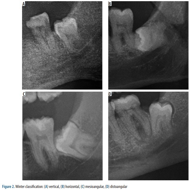
This study analyzed the distribution patterns of impacted mandibular third molars (IMTMs) using the Winter and Pell & Gregory classification systems, which guide surgical planning and complication risk assessment. Additionally, the spatial relationship between IMTMs and the inferior alveolar canal (IAC) was evaluated with cone-beam computed tomography (CBCT) to enhance preoperative risk stratification.
Material and methods:
A total of 1594 IMTMs were retrospectively reviewed in patients with available panoramic and CBCT imaging. Angulation was classified according to Winter, while depth and ramus relation were categorized using Pell & Gregory. CBCT was used to assess IAC relationships. Associations between classifications and IAC contact were analyzed, and logistic regression identified independent risk factors.
Results:
Mesioangular impaction was most frequent (37.6%), and Class II Level B was the predominant Pell & Gregory type (29.6%). CBCT showed IAC contact in 83.5% of cases, most commonly superior and buccal. Logistic regression revealed horizontal impactions had significantly lower odds of contact (OR=0.57, p=0.005), while deep types such as Class III Level C strongly predicted contact (OR=10.22, p<0.001). A “between roots” IAC configuration, undetectable on panoramic radiographs, was noted in 7.5%.
Conclusions:
While Winter classification offers moderate predictive value, Pell & Gregory-particularly deeper and more posterior subtypes-correlates strongly with IAC involvement. Findings highlight the value of CBCT in high-risk IMTM cases to improve radiographic assessment of the canal–root relationship and surgical planning.
This study analyzed the distribution patterns of impacted mandibular third molars (IMTMs) using the Winter and Pell & Gregory classification systems, which guide surgical planning and complication risk assessment. Additionally, the spatial relationship between IMTMs and the inferior alveolar canal (IAC) was evaluated with cone-beam computed tomography (CBCT) to enhance preoperative risk stratification.
Material and methods:
A total of 1594 IMTMs were retrospectively reviewed in patients with available panoramic and CBCT imaging. Angulation was classified according to Winter, while depth and ramus relation were categorized using Pell & Gregory. CBCT was used to assess IAC relationships. Associations between classifications and IAC contact were analyzed, and logistic regression identified independent risk factors.
Results:
Mesioangular impaction was most frequent (37.6%), and Class II Level B was the predominant Pell & Gregory type (29.6%). CBCT showed IAC contact in 83.5% of cases, most commonly superior and buccal. Logistic regression revealed horizontal impactions had significantly lower odds of contact (OR=0.57, p=0.005), while deep types such as Class III Level C strongly predicted contact (OR=10.22, p<0.001). A “between roots” IAC configuration, undetectable on panoramic radiographs, was noted in 7.5%.
Conclusions:
While Winter classification offers moderate predictive value, Pell & Gregory-particularly deeper and more posterior subtypes-correlates strongly with IAC involvement. Findings highlight the value of CBCT in high-risk IMTM cases to improve radiographic assessment of the canal–root relationship and surgical planning.
REFERENCES (23)
1.
Yıldırım H, Büyükgöze-Dindar M. Investigation of the prevalence of impacted third molars and the effects of eruption level and angulation on caries development by panoramic radiographs. Med Oral Patol Oral Cir Bucal 2022; 27: e106-e112. DOI: 10.4317/medoral.25013.
2.
Celikoglu M, Miloglu O, Kazanci F. Frequency of agenesis, impaction, angulation, and related pathologic changes of third molar teeth in orthodontic patients. J Oral Maxillofac Surg 2010; 68: 990-995.
3.
Tantanapornkul W, Okouchi K, Fujiwara Y, Yamashiro M, Maruoka Y, Ohbayashi N, Kurabayashi T. A comparative study of cone-beam computed tomography and conventional panoramic radiography in assessing the topographic relationship between the mandibular canal and impacted third molars. Oral Surg Oral Med Oral Pathol Oral Radiol Endod 2007; 103: 253-259.
4.
Patel PS, Shah JS, Dudhia BB, Butala PB, Jani YV, Macwan RS. Comparison of panoramic radiograph and cone beam computed tomography findings for impacted mandibular third molar root and inferior alveolar nerve canal relation. Indian J Dent Res 2020; 31: 91-102.
5.
Santos KK, Lages FS, Maciel CAB, Glória JCR, Douglas-de-Oliveira DW. Prevalence of mandibular third molars according to the Pell & Gregory and Winter classifications. J Maxillofac Oral Surg 2022; 21: 627-633.
6.
Soman S, Managutti A, Sood R, Rajasekhar R, Anroop A. Comparative radiographic assessment of panoramic and segmented cone beam computed tomographic images of impacted mandibular third molars. World J Dent 2025; 15: 972-978.
7.
Yang Y, Bao DY, Ni C, Li Z. Three-dimensional positional relationship between impacted mandibular third molars and the mandibular canal. BMC Oral Health 2023; 23: 831. DOI: 10.1186/s12903-023-03548-0.
8.
Pogorzelska A, Mielczarek A, Regulski PA, Lee M, Szopinski K. Evaluation of the inferior alveolar canal course in relation to fully and partially impacted lower third molar roots via cone-beam computed tomography. Cureus 2024; 16: e74021. doi: 10.7759/cureus.74021.
9.
Khojastepour L, Khaghaninejad MS, Hasanshahi R, Forghani M, Ahrari F. Does the Winter or Pell and Gregory classification system indicate the apical position of impacted mandibular third molars? J Oral Maxillofac Surg 2019; 77: 2222.e1-2222.e9. DOI: 10.1016/j.joms.2019.06.004.
10.
Bell GW. Use of dental panoramic tomographs to predict the relation between mandibular third molar teeth and the inferior alveolar nerve. Radiological and surgical findings, and clinical outcome. Br J Oral Maxillofac Surg 2004; 42: 21-27.
11.
Sant’Ana LF, Giglio FP, Ferreira O Jr, Sant’ana E, Capelozza AL. Clinical evaluation of the effects of radiographic distortion on the position and classification of mandibular third molars. Dentomaxillofac Radiol 2005; 34: 96-101.
12.
Landis JR, Koch GG. The measurement of observer agreement for categorical data. Biometrics 1977; 33: 159-174.
13.
Shaari RB, Awang Nawi MA, Khaleel AK, AlRifai AS. Prevalence and pattern of third molars impaction: a retrospective radiographic study. J Adv Pharm Technol Res 2023; 14: 46-50.
14.
Al-Dajani M, Abouonq AO, Almohammadi TA, Alruwaili MK, Alswilem RO, Alzoubi IA. A cohort study of the patterns of third molar impaction in panoramic radiographs in Saudi population. Open Dent J 2017; 11: 648-660.
15.
Vasegh Z, Bakhshaei P, Jahanbani M, Mahmoudi Anzabi R. Evaluation of anatomical relationships in the mandibular third molar region based on its angulation and depth of impaction: a CBCT-based study. Oral Maxillofac Surg 2024; 28: 613-622.
16.
Khouri C, Aoun G, Khouri C, Saade M, Salameh Z, Berberi A. Evaluation of third molar impaction distribution and patterns in a sample of Lebanese population. J Maxillofac Oral Surg 2022; 21: 599-607.
17.
Yunus B, Wulansari DP, Tunnizha BM. Lower third molar impaction based on Pell and Gregory classification: a panoramic study. Syst Rev Pharm 2020; 11: 19-23.
18.
Kumar VR, Yadav P, Kahsu E, Girkar F, Chakraborty R. Prevalence and pattern of mandibular third molar impaction in Eritrean population: a retrospective study. J Contemp Dent Pract 2017; 18: 100-106.
19.
Urechescu H, Banu A, Pricop M, Streian F, Pricop A, Cuzic C. The positional relationship between the mandibular canal and the lower third molar determined on cone-beam computed tomography. Medicina (Kaunas) 2025; 61: 1291. DOI: https://doi.org/10.3390/medici....
20.
Neves FS, Souza TC, Almeida SM, Haiter-Neto F, Freitas DQ, Bóscolo FN. Correlation of panoramic radiography and cone beam CT findings in the assessment of the relationship between impacted mandibular third molars and the mandibular canal. Dentomaxillofac Radiol 2012; 41: 553-557.
21.
Prados-Frutos JC, Salinas-Goodier C, Manchón Á, Rojo R. Anterior loop of the mental nerve, mental foramen and incisive nerve emergency: tridimensional assessment and surgical applications. Surg Radiol Anat 2017; 39: 169-175.
22.
Yilmaz S, Adisen MZ, Misirlioglu M, Yorubulut S. Assessment of third molar impaction pattern and associated clinical symptoms in a central Anatolian Turkish population. Med Princ Pract 2016; 25: 169-175.
23.
De Oliveira Gamba T, Alves MC, Haiter-Neto F. Analysis of sexual dimorphism by locating the mandibular canal in images of cone-beam computed tomography. J Forensic Radiol Imaging 2014; 2: 72-76.
Share
RELATED ARTICLE
| ISSN: | 1899-0967 |

We process personal data collected when visiting the website. The function of obtaining information about users and their behavior is carried out by voluntarily entered information in forms and saving cookies in end devices. Data, including cookies, are used to provide services, improve the user experience and to analyze the traffic in accordance with the Privacy policy. Data are also collected and processed by Google Analytics tool (more).
You can change cookies settings in your browser. Restricted use of cookies in the browser configuration may affect some functionalities of the website.
You can change cookies settings in your browser. Restricted use of cookies in the browser configuration may affect some functionalities of the website.
