

Dear Editor,
Color Doppler ultrasound (CDUS) is a rapid, non-invasive, first-line imaging modality for vascular assessment [1]. However, its diagnostic accuracy can be limited when nonvascular lesions produce misleading flow signals. We report a case of an inguinal lymphatic malformation (LM) that artifactually exhibited a “yin-yang” sign during the Valsalva maneuver, mimicking a pseudoaneurysm.
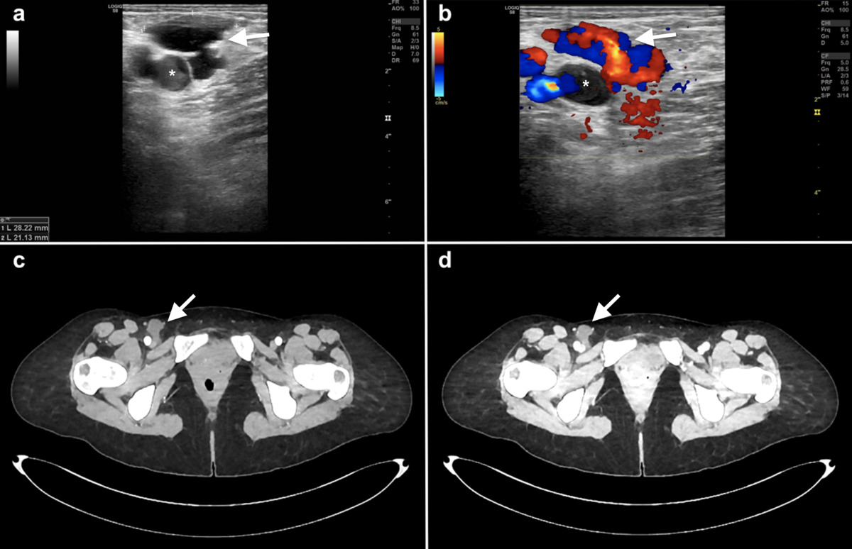
A 46-year-old woman presented with a painless right groin mass. Grayscale ultrasonography revealed a well-defined, anechoic cystic lesion adjacent to the common femoral vein (Figure 1A). CDUS at rest showed no internal flow, but during Valsalva, a “yin-yang” pattern emerged, suggestive of a pseudoaneurysm (Figure 1B). The absence of prior vascular intervention or a characteristic “to-and-fro” spectral waveform raised diagnostic uncertainty. Contrast-enhanced computed tomography confirmed a non-enhancing cystic lesion, excluding a vascular origin (Figure 1C, D). Aspiration yielded serous fluid containing inflammatory cells. The lesion recurred after three weeks, and surgical excision confirmed a macrocystic LM.
Figure 1
Imaging findings of a lymphatic malformation adjacent tothe right common femoral vein. A) Grayscale ultrasound reveals a well-circumscribed, anechoic cystic lesion. B) Color Doppler ultrasound during the Valsalva maneuver demonstrates an artifactual “yin-yang” sign within the cyst, mimicking a pseudoaneurysm. C, D) Axial contrast-enhanced computed tomography images in the (C) arterial and (D) venous phases confirm the non-enhancing cystic nature of the lesion. The arrow indicates the lymphatic malformation. The asterisk (*) denotes the common femoral vein

LMs are congenital, low-flow vascular anomalies resulting from aberrant lymphatic development, most commonly located in the head and neck [2,3]. Inguinal involvement in adults is exceedingly rare [4]. The “yin-yang” sign, typically considered more specific for pseudoaneurysms, was artifactually produced in this case. We suggest that Valsalva-induced pressure changes within the adjacent common femoral vein generated turbulent motion of cystic fluid, creating a pseudo-flow signal on Doppler.
In conclusion, while CDUS remains an invaluable tool for vascular evaluation, the appearance of a “yin-yang” pattern adjacent to a major vein during the Valsalva maneuver – especially in patients without a history of vascular intervention – should prompt careful reconsideration. Awareness of potential mimickers such as LMs is essential to avoid diagnostic errors. When imaging findings are atypical, complementary cross-sectional modalities can play a crucial role in confirming the diagnosis and guiding appropriate management.