


Introduction
Five to fifteen percent of couples worldwide are currently infertile, with female factors accounting for about 65% of cases [1]. Infections, endometriosis, prior surgery, and, in rare cases, fibromas or polyps, are the main causes of tubal blockage, which accounts for 26% of female infertility [2]; 40% of female infertility cases are diagnosed with hysterosalpingography (HSG) or laparoscopic procedures, which are reported to be as accurate in determining uterine morphology [3].
Laparoscopy with dye chromopertubation – commonly known as the lap-and-dye test – is considered the gold standard for evaluating tubal patency. Despite its diagnostic accuracy, it is seldom used due to its invasive nature and high cost. As a result, HSG remains the most widely adopted method across many countries. HSG is recognized for being accurate, safe, efficient, and cost-effective. However, it comes with notable drawbacks that prompt the search for alternatives: it can be painful, involves the use of iodinated contrast agents, and exposes patients to ionizing radiation [4]. Transvaginal ultrasound alone cannot determine whether the fallopian tubes are patent. In contrast, hysterosalpingo contrast sonography (HyCoSy) is recognized as a sensitive and efficient method for evaluating tubal patency. It offers a quick, straightforward, and dependable screening process while simultaneously allowing assessment of the uterus and ovaries [5]. HyCoSy has been considered as a less painful alternative to HSG; it is a minimally invasive ultrasound-based procedure, typically performed by gynecologists, that eliminates the risks associated with iodinated contrast agents and exposure to ionizing radiation [2]. It involves transvaginal ultrasound-aided intrauterine contrast administration, allowing assessment of tubal patency and identification of gynecological conditions such as endometrial polyps, fibroids, and ovarian cysts, and evaluation of endometrial thickness [6]. Early contrast agents used in the procedure included saline or a mixture of air and saline. However, interpreting tubal patency with these agents required advanced skill due to the brief window during which the contrast could be visualized passing through the fallopian tubes [7]. Then, more echogenic contrast media such as Echovist Infoson and SonoVue were introduced, but they are either no longer marketed or not authorized for intrafallopian usage [8]. ExEm gel (GynaecologIQ), made of glycerol and hydroxyethylcellulose, was released in 2007. It is diluted with water, and when this solution is forced through small holes in tubes and syringes, foam is produced mechanically. In comparison to water, the diluted gel is sufficiently fluid to flow through patent tubes without being so viscous that the air bubbles stay suspended in it for an extended period. As a result, the foam mixture becomes more stable and can continue to be echogenic for roughly seven minutes, making it suitable for use as a contrast medium in hysterosalpingo-foam sonography (HyFoSy) [9].
Although many studies have shown comparable efficacy between HyFoSy and HSG, a critical knowledge gap remains in the current evidence [10,11].
There is no standardized protocol for bias estimation, and current methods do not adequately account for patient-specific factors, such as anatomical variations, that affect diagnostic concordance. The data on false positive HyFoSy findings are scarce, limiting evidence-based recommendations for routine test selection practice [12].
Furthermore, there is a lack of comparative data from Iraq, underscoring the need for regional validation to establish baseline diagnostic parameters.
This study aimed to assess the diagnostic agreement between HyFoSy and HSG as a test for tubal patency assessment via prevalence-adjusted statistical methods to define systematic bias. The secondary aim was to determine the impact of patent criteria, specifically the position of the uterus, on test concordance, to build a permanent regional dataset for future validation. A deeper insight into the magnitude and the direction of diagnostic concordance between the two tests is crucial for developing an evidence-based clinical algorithm that balances diagnostic accuracy with patient comfort.
Material and methods
Participants and ethics
This prospective comparative diagnostic accuracy study was performed in the Radiology Department of Al-Yarmouk Teaching Hospital for 5 months (from March to July, 2025). The Ethical Committee of Al-Kindy College of Medicine issued the study approval No. 217 in 30/04/2025. All the study methods were conducted in line with the Declaration of Helsinki. Written informed consent was obtained from all participants after they were informed of the study’s aim, procedure, and potential radiation risks. All participants were informed that they had the right to withdraw without compromising their right to clinical care.
Consecutive sampling was used to enroll infertile women referred for a traditional HSG examination as part of an infertility workup. Patients who consented to undergo both examinations (HSG and HyFoSy) were included in the study.
Inclusion criteria
Infertile female patients aged from 18 to 45 years old were invited; infertility was defined as failure to achieve pregnancy following 1 year of regular unprotected sex (reduced to 6 months for women older than 35 years) of regular unprotected sex [13].
Clinical indication for tubal patency assessment.
The patient should have documented normal ovulation, although HSG is often performed in women with concurrent ovulatory problems in practice; we adopted this criterion to create a more homogeneous population in which infertility is attributable primarily to tubal factors.
Regular baseline renal function test; creatinine < 1.2 mg/dl.
Exclusion criteria
Participants with a history of contrast allergic reaction or intolerance to one of the two examinations were excluded from the study.
Active pelvic inflammatory disease or severe systemic illness.
Unexplained abnormal uterine bleeding.
Both tubal patency assessments were conducted within two weeks from the first day of menstruation, preferably between days 6 and 10.
Operator qualification and blindness, outcome measured
The physician conducting the test was a certified radiologist with more than 5 years of expertise in reproductive imaging in the presence of a gynecologist. The radiologist conducting the HSG was unaware of the HyFoSy results, and likewise, the HyFoSy examiner was blinded to the HSG findings. The outcomes of both tests were categorized as normal, unilateral tubal pathology, or bilateral tubal pathology.
Imaging protocol details
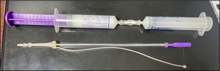
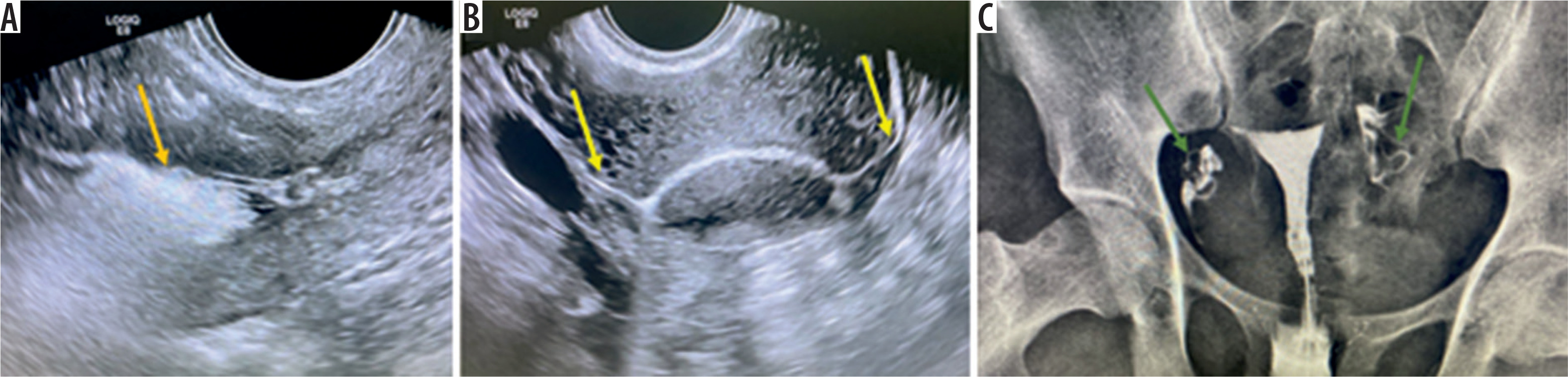
Beginning with the HyFoSy examination, the patient is put in a lithotomy position, and after sterilization of the genital area, a suitable size Cusco’s speculum is introduced to visualize the vagina and cervical canal. A small cervical catheter is introduced through the cervix and connected to a syringe containing 10 cc of foam, which is then injected into the uterus (Figure 1). A foam-filled syringe is attached to this applicator. In a 10 ml syringe, 5 ml of pure water and 5 ml of ExEm-gel (IQ Medical Ventures BV, Delft, The Netherlands; containing hydroxyethyl cellulose and glycerol) are thoroughly mixed to generate this foam, producing foam that is stable enough to exhibit echogenicity for at least five minutes and that has enough fluid to flow through patent tubes. Transvaginal ultrasound is performed to determine whether the fallopian tubes are patent during the foam infusion into the uterus. This foam material appears highly echogenic, outlining the endometrial cavity, one or both fallopian tubes, and spills within the pelvic cavity (Figure 2).
During the HSG examination, a balloon catheter is placed on the cervix, which is used to inject roughly 10 cc of water-soluble iodinated contrast medium into the uterus and fallopian tubes, depending on the local protocol of our hospital. The flow of the contrast medium within the uterus and the tubes can be visualized by exposing the pelvic area to a series of about six radiographs to determine their patency while the contrast medium is inserted into the uterus and fallopian tubes (Figure 2).
Both examinations (HSG and HyFoSy) were conducted in random order throughout the follicular phase of the same menstrual cycle (5th to 11th cycle day). The physicians were blinded to the results of the previously performed examination, according to the gold standard FOAM study protocol, which stated that conducting both tests within the same cycle does not impact the visible tubal patency compared with each method alone [14].
Sample size calculation
The sample size was determined according to prior studies, which reported agreement rates between the HyFoSy and HSG tubal patency tests [15,16].
Figure 1
Two syringes of foam and water (orange arrows) connected by a connector (green arrow) for mixing. The cervical applicator (blue arrow) is used to introduce the material to the cervix and uterus
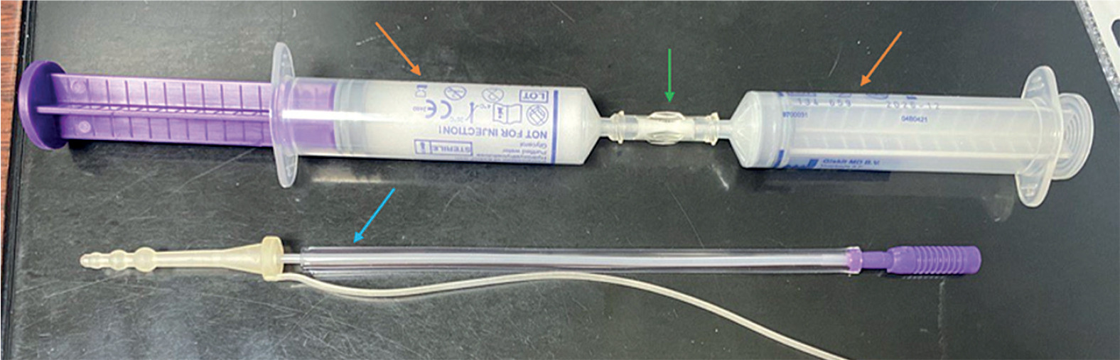
Figure 2
Transvaginal ultrasound images after injection of the foam into the uterus. A) Echogenic foam material outlining the endometrial cavity (orange arrow). B) Spillage of ultrasonic contrast in both patent tubes (yellow arrows). C) Hysterosalpingography of the same patient showing patency of both tubes (green arrows)
A Cohen’s kappa (κ) coefficient of 0.7 was targeted, which is interpreted as substantial agreement compared to the null hypothesis of 0.4, interpreted as moderate agreement [17]. With an α value of 0.05 and a statistical power of 80%, the calculation yielded a required sample size of 72 participants. To address potential technical failure, patient withdrawal, and subgroup analysis, we opted to enroll 80 participants.
Statistical methods
Data were analyzed by SPSS 26, and baseline criteria were presented as counts and percentages. Fisher’s exact test was used to examine the association between categorical variables and diagnostic concordance, given the small sample size. We evaluated the diagnostic agreement between HyFoSy and HSG using the overall agreement percentage, Cohen’s k coefficient for chance-corrected agreement, and Gwet’s agreement coefficient 1 (AC1) to adjust for prevalence bias. McNemar’s test examined systematic differences between the two paired methods. Positive and negative agreement proportions were added to the calculations. A Bland-Altman analysis evaluated agreement across the ranges of the tubal patency score. A p < 0.05 was set as significant for all tests.
Results
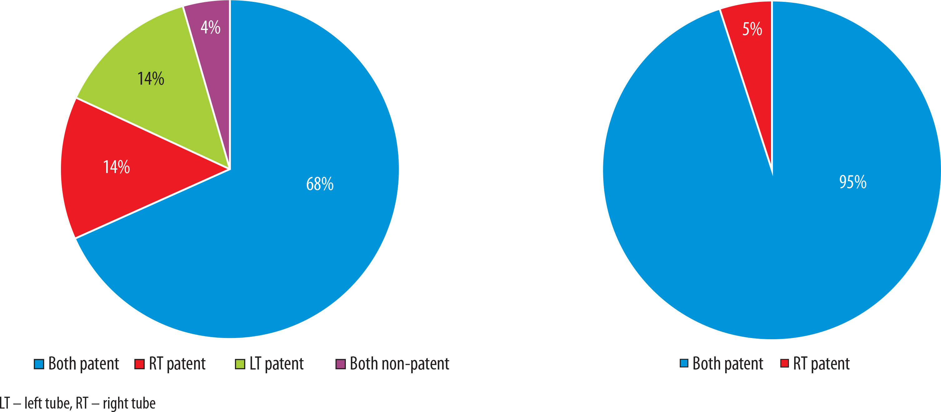
A total of 80 women of childbearing age were included in this study, with the majority, 64 (80%), aged less than 35 years, and 48 (60%) were nulliparous. The uterine position was anteverted in 64 (80%) vs. 16 (20%) that were retroverted in the included sample. HyFoSy shows high diagnostic concordance when compared to HSG, with concordance demonstrated in 68 (85%) women, as shown in Table 1 and Figure 3.
Table 1
Clinical and anatomical characteristics of the studied sample (N = 80)
| Variable | n (%) |
|---|---|
| Age | |
| ≤ 35 years | 64 (80) |
| > 35 years | 16 (20) |
| Parity | |
| Yes | 32 (40) |
| No | 48 (60) |
| Uterus | |
| AV | 64 (80) |
| RV | 16 (20) |
| Diagnostic accuracy | |
| Concordance | 68 (85) |
| Discordance | 12 (15) |
Figure 3
Diagnostic concordance between (A) hysterosalpingo-foam sonography (HyFoSy) and (B) hysterosalpingography (HSG) in tubal patency
Overall, HyFoSy and HSG showed concordant findings in 85% of the patients. Uterine position significantly affected the agreement rates (p = 0.028). Anteverted (AV) uterus showed a meaningfully high concordance (88.2%) vs. retroverted (RV) uterus (33.3%). Age and parity showed no significant associations (Table 2).
Table 2
Diagnostic agreement between hysterosalpingo-foam sonography (HyFoSy) and hysterosalpingography (HSG) by patient characteristics
| Variables | Diagnostic concordance (n = 68) | Diagnostic discordance (n = 12) | Odds ratio (95% CI) | p-value (Fisher’s exact test) |
|---|---|---|---|---|
| Age, n (%) | ||||
| < 35 years | 56 (82.4) | 8 (66.7) | 2.09 (0.65-6.74) | 0.509 |
| > 35 years | 12 (17.6) | 4 (33.3) | Reference | |
| Parity, n (%) | ||||
| Yes | 28 (41.2) | 4 (33.3) | 1.38 (0.38-4.94) | 0.998 |
| No | 40 (58.8) | 8 (66.7) | Reference | |
| Uterus position, n (%) | ||||
| AV | 60 (88.2) | 4 (33.3) | 15.0 (3.67-61.37) | 0.028* |
| RV | 8 (11.8) | 8 (66.7) | Reference | |
Both methods agreed on bilateral tubal patency in 70% of the cases. Interestingly, no bilateral non-patent cases were diagnosed by either method, which hinders the assessment of negative agreement (Table 3).
Table 3
Cross-tabulation of tubal patency findings between the two methods (per-patient analysis, N = 80)
| HyFoSy results | HSG: both patent, n (%) | HSG: unilateral patent, n (%) | HSG: both non-patent, n (%) | Total, N (%) |
|---|---|---|---|---|
| Both patent | 56 (70.0)* | 16 (20.0)† | 4 (5.0)† | 76 (95.0) |
| Unilateral patent | 4 (5.0)† | 0 (0.0) | 0 (0.0) | 4 (5.0) |
| Both non-patent | 0 (0.0) | 0 (0.0) | 0 (0.0) | 0 (0.0) |
| Total | 60 (75.0) | 16 (20.0) | 4 (5.0) | 80 (100.0) |
The measurement of agreement showed an overall 85% agreement between the two methods. Cohen’s κ was 0.3 (95% CI: 0.05-0.55), indicating fair agreement and suggesting an influence of the low prevalence of negative findings. Gwet’s AC1 coefficient was 0.84, indicating strong agreement (95% CI: 0.74-0.94) after adjusting for prevalence bias. McNemar’s test confirmed a significant systematic bias between the two methods (p < 0.001), suggesting that the interchangeability of the two methods has no role (Table 4).
Table 4
Statistical measure of agreement between hysterosalpingo-foam sonography (HyFoSy) and hysterosalpingography (HSG) (per-tube analysis, N = 160 tubes)
[i] Note 1. Agreement statistics are based on per-tube analysis (160 tubes from 80 patients). Each tube was classified as either patent or non-patent. The overall agreement of 85% indicates that 136 out of 160 tubes (68 out of 80 patients with bilateral agreement) showed concordant results between HyFoSy and HSG.
The Bland-Altman plot showed a mean bias of +0.25 with HyFoSy, with a tendency to overestimate tubal patency versus HSG. Agreement’s limits were –0.98 to +1.48, implying substantial inter-method variability, which suggests that both methods yield nonequivalent measures (Figure 4).
Figure 4
Bland-Altman plot; hysterosalpingo-foam sonography (HyFoSy) vs. hysterosalpingography (HSG) tubal patency score. Blue star markers represent individual paired observations, with each point indicating agreement between HyFoSy and HSG tubal patency scores for a single assessment. The vertical position reflects the difference between the methods, while the horizontal position represents their mean value
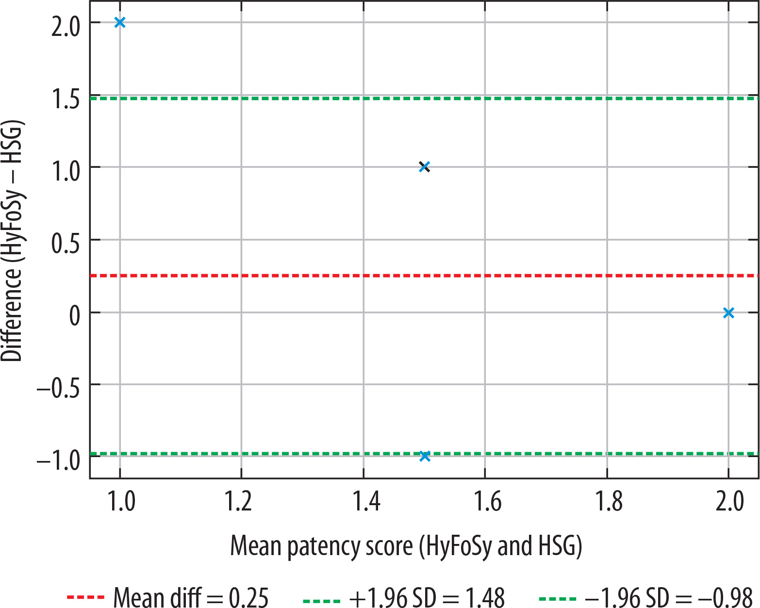
Table 3 provides the raw cross-tabulation of findings on a per-patient basis, showing how the bilateral tubal status classifications compare between the two methods. Table 4 presents statistical agreement measures using per-tube analysis, where each of the 160 tubes (from 80 patients) is evaluated independently for patency. Table 5 presents diagnostic test performance using per-patient analysis, specifically evaluating HyFoSy’s ability to detect bilateral tubal patency when compared to HSG as the reference standard.
Table 5
Summary of diagnostic performance of hysterosalpingo-foam sonography (HyFoSy) compared to hysterosalpingography (HSG) in detecting tubal patency (per-patient analysis, N = 80)
| Parameter | True positive | False positive | True negative | False negative | Sensitivity | Specificity | PPV | NPV | Accuracy |
|---|---|---|---|---|---|---|---|---|---|
| HyFoSy vs. HSG | 56 | 20 | 0 | 4 | 93.3% | 0%* | 73.7% | 0%* | 70.0% |
The different units of analysis (per tube vs. per patient) explain why the total agreement in Table 4 (68/80 patients = 85%) differs from the distribution shown in Table 5, where only cases with bilateral patency agreement (56 patients) are counted as true positives.
Discussion
Tubal patency assessment is a cornerstone in infertility workups. While HSG is considered the gold standard, HyFoSy has emerged as a promising alternative, offering comparable diagnostic accuracy with greater patient comfort and procedural simplicity [6]. ExEm gel is safe, with no adverse effect on blastocyst development [4]. Unlike laparoscopy, which carries inherent risks associated with surgery and anesthesia, or HSG, which uses iodinated contrast agents that may trigger allergic reactions, in addition to radiation risk, HyFoSy provides a safer, less invasive alternative [2].
This study examined the overall concordance between HyFoSy and HSG in testing tubal patency, revealing a high concordance rate of 85% and a Cohen’s κ of 0.30 with a 95% CI of 0.05 to 0.55. This finding was further confirmed by Gwet’s AC1, which indicated strong agreement (0.84). In evaluating tubal patency, HyFoSy and HSG have a fair agreement. These results align with findings from studies by van Welie et al. [14], Emanuel et al. [17], Tsakos et al. [10], and Ramos et al. [15], as presented in Table 6.
Table 6
Agreement between hysterosalpingo-foam sonography (HyFoSy) and hysterosalpingography (HSG) according to previous studies
| Study/year | Study type, No. of participants | Agreement | Interpretation |
|---|---|---|---|
| Van Welie et al. (2022) [14] | RCT, 1026 | ~84% | HyFoSy was non-inferior to HSG in guiding fertility management; however, discordance was noted in ~16% of cases |
| Emanuel et al. (2011) [17] | Prospective observational cohort study, 72 | 78% | Discordance in 7% of cases; HyFoSy avoided HSG in most patients, showing good feasibility |
| Tsakos et al. (2023) [10] | Systemic review and meta-analysis, 1433 | Fair agreement The analysis showed a Cohen’s κ value of 0.39 | Meta-analysis showed HyFoSy was less painful and more reliable than the HSG |
| Ramos et al.(2021) [15] | Prospective observational cohort study, 106 | Moderate agreement HyFoSy showed total concordance with HSG in 72.6% of cases and total discordance in 4.7% Cohen’s κ = 0.57 | HyFoSy is a useful method for determining tubal patency; it might be regarded as a viable substitute for HSG in terms of precision and efficacy in patients with minimal tubal disease risk |
The analysis showed that the HyFoSy and HSG test reliability was not affected by patient age or parity; both showed no influence on the agreement, in line with earlier works [11,15].
Li et al. [18] reported no significant correlation between primary or secondary infertility regarding fallopian tube visualization. A woman’s status as multiparous or nulliparous had no bearing on the procedure’s success, tolerability, or diagnostic performance. This could indicate that parity has little bearing on contrast flow, tubal visualization, or patient discomfort during HyFoSy.
The current results confirmed that the test agreement was reduced in RV uteri to 33%, with a p-value of 0.001, which underscores the uterine position as a key determinant in the accuracy of the HyFoSy test.
This may be due to many factors. For example, a uterus that is sharply retroverted may make cervical catheterization more difficult and raise the possibility of less than ideal contrast flow. The retroverted uterus also affects the trajectory of foam contrast, which may result in false negatives or poor fallopian tube visualization, so to see the tubes clearly in retroverted uteri, the sonographer may need to make significant adjustments to the probe orientation [19].
Li et al. [18] stated that the uterine position, uterine angle, and ovarian position all affect the visualization of the fallopian tubes, in good agreement with our results. Bohîlțea et al. [12] recommended a wedge-shaped pad or a special gynecological table to ensure a gynecological position that is as close to 45o as possible to the Trendelenburg position to reorient the fallopian tubes as anatomically as possible.
Overestimation of tubal patency has clinical implications: the patient will experience delayed referral for definitive diagnosis and undergo a misguided treatment strategy that will only prolong the infertility period, with associated consequences [20].
We found that bilateral tubal patency in both examinations was 70% (perfect agreement) of cases in both HSG and HyFoSy. In comparison, Exacoustos et al. [21] observed 81% bilateral patent tubes. This disparity could be due to the use of 3D ultrasound compared with the 2D ultrasound we used.
The Bland-Altman plot shows a mean bias of +0.25 and wide limits of agreement (–0.98 to +1.48), suggesting that HyFoSy tends to overestimate tubal patency compared to HSG, and the variability between methods is substantial enough to question their interchangeability.
The study results of Ramos et al. [15] and Grigovich et al. [22] aligned with ours; they stated that HSG remains the reference standard due to its ability to detect subtle tubal pathology, while HyFoSy is more patient-friendly but may miss certain structural abnormalities that HSG can detect, contributing to inter-method variability.
Several factors may explain why HyFoSy foam tends to overestimate tubal patency; the foam is compressible, so it may pass through partially obstructed tubes and give a false positive for a patent tube. The pressure used to inject the foam exacerbates this; it may increase its passage through anatomically patent tubes [23,24].
However, in reality, the tubes are physiologically obstructed by adhesions, so the picture obtained is not a reflection of what really happens in normal physiology, where these adhesions impede ciliary movement of sperm; i.e., HyFoSy may miss tubal impaired ciliary movement or mucosal damage [25].
Moreover, technical issues play a role, as the ultrasonography examination is operator-dependent and prone to acoustic shadowing, which may partially mask a blocked tube diagnosed by HSG during an X-ray examination [26-28].
The study showed a good overall agreement between the two tests and a strong statistical concordance. Critically, McNemar’s test revealed systematic bias, which indicated that both tests have strong overall concordance, but they are not interchangeable. This implies that in high-risk groups or in circumstances where definitive tubal assessment is needed to shape a patient treatment plan, HSG may serve as a better tool due to false negative results.
HyFoSy is a useful non-invasive first-line screening tool in low-risk infertile women with AV uteri. However, in RV uteri, a confirmatory HSG remains essential. Clinicians should discuss HyFoSy limitations, and a comprehensive, integrated anatomical-functional evaluation is advised to reduce the odds of false reassurance, and delayed appropriate management, thereby aiming to optimize patient-centered care.
Limitations
We acknowledge the study’s limitations, including its small sample size, which limits the generalizability of its results, and the absence of cases with bilateral tubal blockage, which restricted the evaluation of negative agreement. Finally, the potential technical challenges with RV uteri may have affected the diagnostic accuracy.
Our inclusion criterion requiring normal ovulation may limit generalizability, as tubal patency assessment is often performed in women with concurrent ovulatory disorders in clinical practice.
Strengths
The prospective study design and well-defined inclusion criteria strengthen the study’s internal validity. The prevalence bias was addressed by multiple statistical agreement tests (κ, Gwet’s AC1). Being conducted in a real-world clinical setting enhances the study’s external validity.
Suggested future works
Larger multicenter studies with a larger sample size are needed to confirm these findings and to refine imaging techniques for RV uteri. HyFoSy may ultimately be integrated with other diagnostic fertility tests as part of a comprehensive infertility workup.
Conclusions
The substantial diagnostic discrepancies between HyFoSy and HSG make HyFoSy a valuable complementary tool for infertility workup, owing to its ease of administration and non-invasive nature. However, the systematic tendency of HyFoSy to overestimate tubal patency, especially among women with RV uterus, highlights the need for careful patient stratification according to the uterus anatomy. A tailored diagnostic approach, using HyFoSy as the first-line screening in women with AV uteri, is recommended, whereas direct referral to HSG is advised for RV uteri or when a definitive assessment is crucial for guiding treatment strategy. These findings should be interpreted with caution, given the small sample size and the lack of bilateral tubal blockage cases in our cohort, which warrants larger studies with a more balanced case distribution to validate the current results.

