

Introduction
Allogeneic hematopoietic cell transplantation (alloHCT) is a lifesaving procedure for several hematological, otherwise incurable diseases. However, there is a high risk of procedure-related complications. One of the most common complications is kidney injury. The etiology of kidney injury in this group of patients is complex and involves the effects of nephrotoxic drugs, infectious complications, microangiopathy, cytokine release syndrome, sinusoidal obstructive syndrome/veno-occlusive disease (SOS/VOD) and, potentially, graft-versus-host disease (GvHD) [1-4]. Acute kidney injury (AKI) negatively impacts survival outcomes [1,5].
The routine diagnostic workup of kidney injury includes serum creatinine concentration, urine examination, and imaging studies, most commonly ultrasound, including a Doppler-based method. Whenever the cause of kidney dysfunction needs to be determined, kidney biopsy is performed. This, however, is frequently contraindicated in the field of alloHCT due to a low platelet count or impaired coagulation. Therefore, novel strategies are needed in this vulnerable population. These involve new kidney injury biomarkers of serum and urine [1] and assessment of the renal resistive index (rRI) determined by Doppler-based ultrasound and kidney elastography. This study evaluated the role of rRI and renal elastography in the field of alloHCT.
The rRI reflects intrarenal artery resistance. It is a potential marker of glomerulosclerosis, arteriolosclerosis, and tubulointerstitial damage as well as AKI [6] or chronic kidney disease (CKD) [7]. Increased values of rRI have a potential to predict prognosis and responsiveness to steroid therapy in patients with CKD [8].
Elastography is a rapidly developing non-invasive technique that quantitatively measures tissue elasticity by measuring shear wave velocity. It uses acoustic pushing pulses to record the tissue’s dynamic response. Higher shear wave velocity values indicate higher stiffness of the tissue [9]. Elastography is already used in the diagnosis of liver fibrosis and cirrhosis, as well as tumors of the breast and thyroid gland, and is widely investigated in the field of kidney injury [10]. However, the complex anatomy and location of the kidney make it more challenging to assess its elasticity. Several methods, including acoustic radiation force impulse imaging, shear wave elastography (SWE), and Young’s modulus, have been developed to increase the accuracy of kidney elastography [11]. SWE is particularly practical since it can be performed with a conventional probe. However, the results of kidney elastography are variable, which makes it unavailable for routine practice [12]. Nevertheless, it is developing as a tool for transplanted kidney monitoring [12,13]. In the field of alloHCT, liver elastography is potentially useful to establish the diagnosis of SOS/VOD [14].
To our knowledge, neither rRI nor elastography of the kidney has been evaluated in the field of alloHCT. The aim of this pilot observational study was to assess the utility of rRI and renal SWE (rSWE) in evaluating the risk of kidney injury, describing its fluctuations, and determining its influencing factors. Our main hypothesis was that rRI and rSWE are associated with the risk of kidney dysfunction beyond 100 days after alloHCT and that the values change after alloHCT, corresponding to significant complications of alloHCT: sepsis, AKI and GvHD. The objectives were to compare the baseline values in alloHCT recipients with the control group and correlate them with renal function tests. Moreover, repeated measurements and their ratios to baseline were compared between groups with sepsis, AKI, and acute and chronic GvHD.
Material and methods
Study population
We enrolled 33 adult patients eligible for alloHCT in the Department of Hematology, Transplantation and Internal Medicine of the Medical University for Warsaw between April 2019 and April 2022 and agreed to undergo Doppler-based ultrasound and rSWE at three time points: before conditioning, at the time of discharge from hospital, and at least 100 days after the procedure. The recruitment was suspended in May 2020 due to COVID-19 pandemic regulations and resumed in February 2021. Clinical data were gathered from clinical reports prospectively. Routine kidney function tests were performed at least three times a week during hospitalization and weekly after discharge.
Additionally, retrospective data of 30 patients adjusted for sex and age were collected to establish the control group. They were ambulatory patients with normal kidney function and no hematologic diseases who underwent abdominal ultrasound performed for other reasons. The number of subjects in this pilot study was planned based on previous studies that evaluated rRI and rSWE in the field of CKD [6,10,11,15,16].
The study was approved by the institutional review board of the Medical University of Warsaw (No. KB33/2019). Written informed consent was obtained from all patients.
Study end points
The primary end points were rRI and rSWE in the population of alloHCT reciepients and their changes in two repeated measurements: at the time of discharge from hospital and beyond 100 days after alloHCT. Secondary endpoints were differences of rRI and rSWE between alloHCT patients and the control group and factors that potentially influence values of rRI and rSWE in alloHCT reciepients: AKI, sepsis, and GvHD.
Variabilities
Patients received conditioning treatment before alloHCT according to local standards; chemotherapy included busulfan (Bu) or treosulfan with fludarabine (Flu) or cyclophosphamide (Cy) for myeloid malignancies. For lymphoid malignancies, conditioning regimens included etoposide with Cy and total body irradiation (TBI), Flu with melphalan or Bu, and Bu with Cy. Patients with aplastic anemia received reduced intensity conditioning with Cy, Flu, and rabbit anti-thymocyte globulin. The intensity of conditioning was defined according to Center for International Blood and Marrow Transplant Research (CIBMTR) criteria [17]. Rabbit anti-thymocyte globulin was used in transplantations from unrelated donors. GvHD prophylaxis was based on cyclosporin A (CSA) and a short course of methotrexate or CSA and mycophenolate mofetil (MMF). Post-transplant Cy followed by tacrolimus (TAC) and MMF was applied in alloHCT from haploidentical donors. Infection prophylaxis included fluoroquinolone or cephalosporin, acyclovir and micafungin or fluconazole until engraftment followed by penicillin V, acyclovir, fluconazole, and trimethoprim-sulfamethoxazole until cessation of immunosuppression.
Imaging acquisition
Patients were asked to fast for at least four hours and empty the bladder directly before the examination. Conventional and Doppler-based ultrasound and rSWE were performed by the same radiologist (M.J.), using a Toshiba Aplio 500 ultrasound machine (Toshiba Medical Europe, Zoetermeer, The Netherlands) and a Convex 1.9-6 MHz transducer PVT-375BT. Patients were in a supine position. Length and parenchymal thickness of both kidneys were measured. The interlobar arterial resistive index and rSWE were most often taken from the right kidney or the left one if it was more available for visualization. For rSWE, a region of interest sized 10 × 10 mm was positioned in the central part of kidney parenchyma with breath-hold. A mean of 10 measurements was obtained for each subject and expressed in m/s. Given the complex anatomy of the kidney and variability in depths of measurements of native kidneys, the interquartile range (IQR) was considered valid when the value was < 0.5.
Definitions
For the purpose of the study, we used Kidney Disease: Improving Global Outcomes (KDIGO) definitions of AKI and CKD [18,19]. Since the hourly diuresis was not always monitored, the criteria based on diuresis were not included. Glomerular filtration rate (eGFR) was estimated using the Chronic Kidney Disease Epidemiology Collaboration (CKD-EPI) formula [20]. GvHD was defined according to the current National Institutes of Health (NIH), European Society for Blood and Marrow Transplantation (EBMT), and CIBMTR statement [21].
Statistical analysis
The results were analyzed using the statistical software Statistica, version 13.3 (StatSoft Krakow, Poland), and the significance level was set as 0.05. Mean values, standard deviations, medians, and interquartile ranges of measurements were reported. The normality was assessed using the Shapiro-Wilk test. The results were then compared between groups using the independent sample t-test or Mann-Whitney U test as appropriate. The chi-square (c²) and Fisher exact tests were used for the analysis of nominal data. Differences between measurements at three time points were analyzed with one-way analysis of variance (ANOVA). The correlations between rRI and rSWE, renal function tests, and selected clinical outcomes were assessed by Pearson’s correlation coefficient or the Spearman rank correlation test as appropriate.
Results
Characteristics of patients
Sixty-three individuals were recruited to the study (33 patients and 30 controls) and had baseline conventional and Doppler-based ultrasound and rSWE. The median age was 52 years, and 34.4% were female. The baseline characteristics are shown in Table 1.
Table 1
Baseline characteristics of patients (N = 33)
[i] alloHCT – allogeneic hematopoietic cell transplantation, ATG – anti-thymocyte globulin, BMI – body mass index, BSA – body surface area, CR1 – first complete remission, CSA – cyclosporin A, eGFR – estimated glomerular filtration rate, GvHD – graft versus host disease, HCT-CI – hematopoietic cell transplantation comorbidity index, IQR – interquartile range, MMF – mycophenolate, MTX – methotrexate mofetil, sCr – serum creatinine concentration, rRI – renal resistive index, rSWE – renal shear wave elastography, SD – standard deviation, TAC – tacrolimus
Twenty-two patients had repeated examination at discharge (median day +29, range 21-71) and 13 patients were examined again after a median of 105 days following alloHCT (range 93-170). Three patients died before day 100 after alloHCT; one of the deaths was treatment-related mortality (hepatic GvHD). Three patients who relapsed before day +100 were excluded from testing after day +100. The other participants were not fit for measurements due to infection, hospitalization, or lack of compliance. Four rSWE results were considered invalid due to IQR > 0.5 (two baseline, one at discharge, and one after 100 days). At discharge, 16 of the examined patients were on immunosuppressive treatment with CSA, while three were on TAC and one received sirolimus with MMF for severe acute GvHD. At the day of measurement after 100 days, seven patients were receiving CSA, three were receiving TAC, and one remained on sirolimus and MMF.
Within the first 100 days after alloHCT, 15 patients were diagnosed with AKI; three of them were grade two, and none was grade three. Median time to AKI was 34 days (range –8 to +95). Causes of AKI were diverse: calcineurin inhibitors (CNI; 4), TBI (1), cytomegalovirus and BK virus reactivation (1), septic shock (1), other infection (1), and unknown in five cases. Twenty-nine patients survived one year (two deaths attributed to relapse and two non-relapse), among whom six patients developed stage three CKD (eGFR < 60) after one year.
Baseline rRI
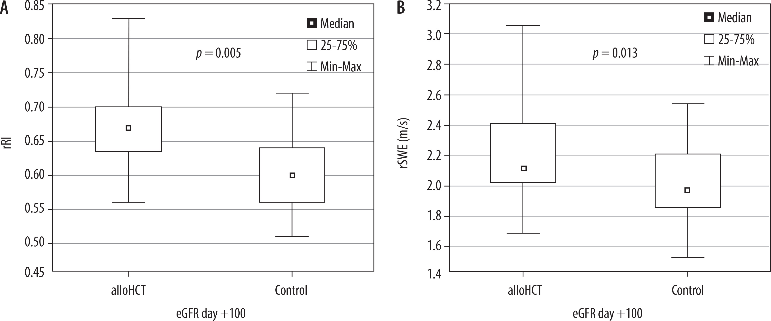
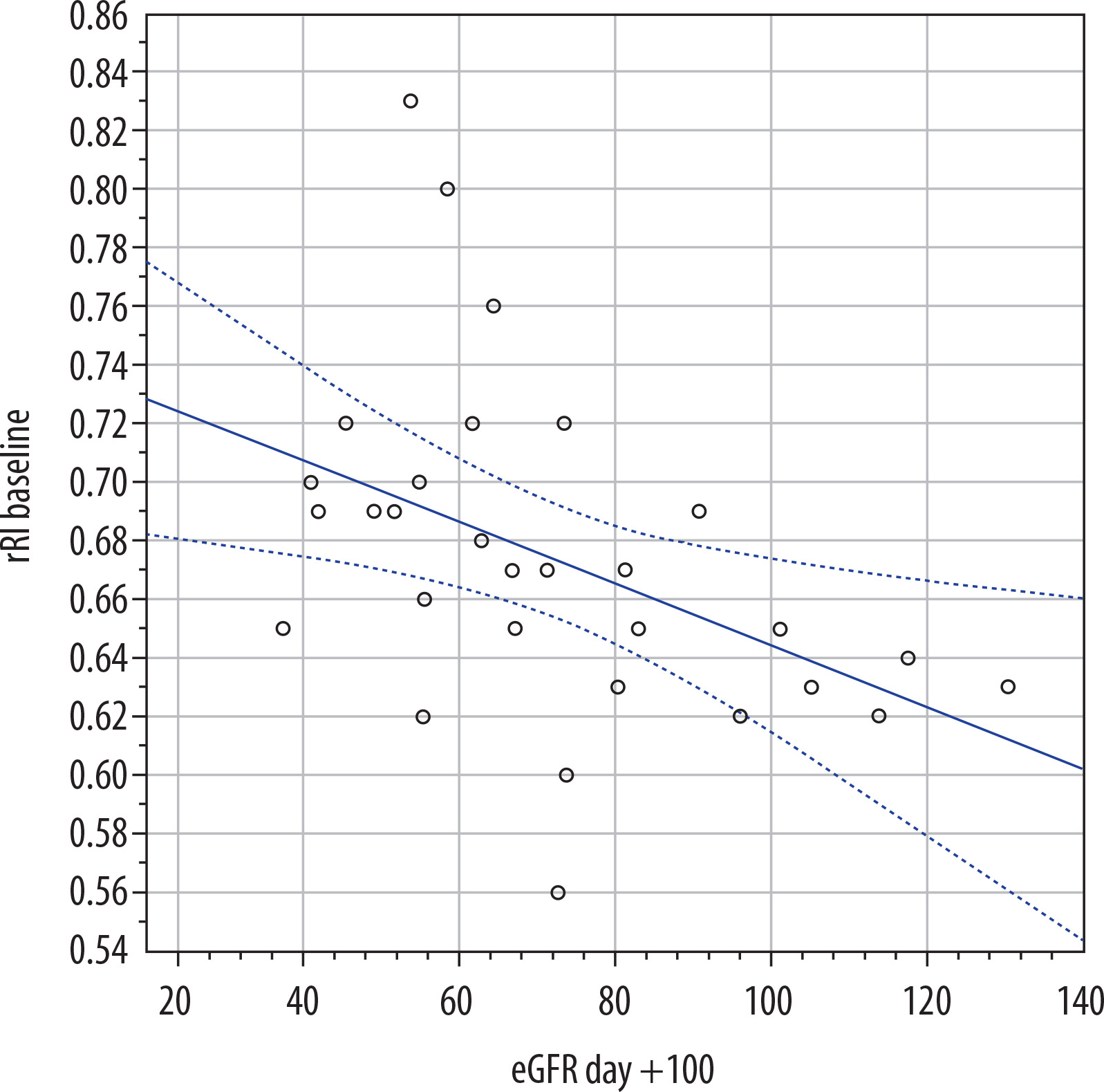
At baseline, mean rRI was 0.68 (IQR 0.65-0.70), which was significantly higher than in the control group (p = 0.005, mean difference 12%; 95% confidence interval [CI]: 5-22%) (Table 2, Figure 1). A positive correlation was found between rRI and age (r = 0.420; 95% CI: 0.08-0.66; p = 0.017) and BMI (r = 0.400; 95% CI: 0.06-0.60; p = 0.023). Baseline serum creatinine concentration and eGFR did not correlate with rRI (Supplementary Table 1), but correlations were found with the measurements after day +100: positive with creatinine after day +100 (r = 0.45; 95% CI: 0.11-0.69; p = 0.012) and negative with eGFR (r = –0.56; 95% CI: 0.25-0.76; p = 0.001, Figure 2).
Table 2
Comparison of patients and control group
Baseline rSWE
Median baseline rSWE was 2.11 (IQR 2.02-2.41), and it was higher than in the control group (p = 0.013, median difference 7%; 95% CI: 2-15%) (Table 2, Figure 1). No correlations were found between rSWE and age, BMI, or renal function tests (Supplementary Table 1).
Repeated measurements
rRI
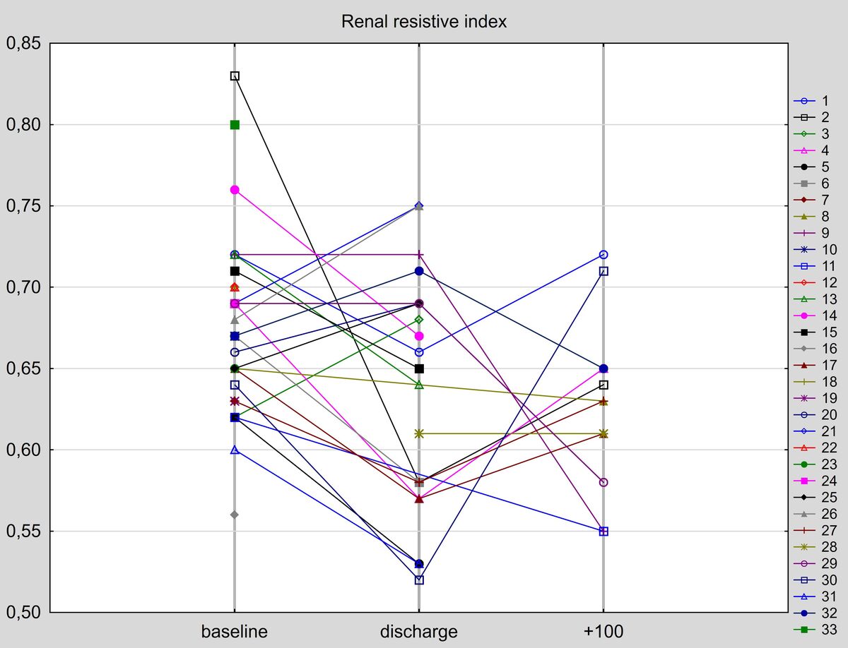
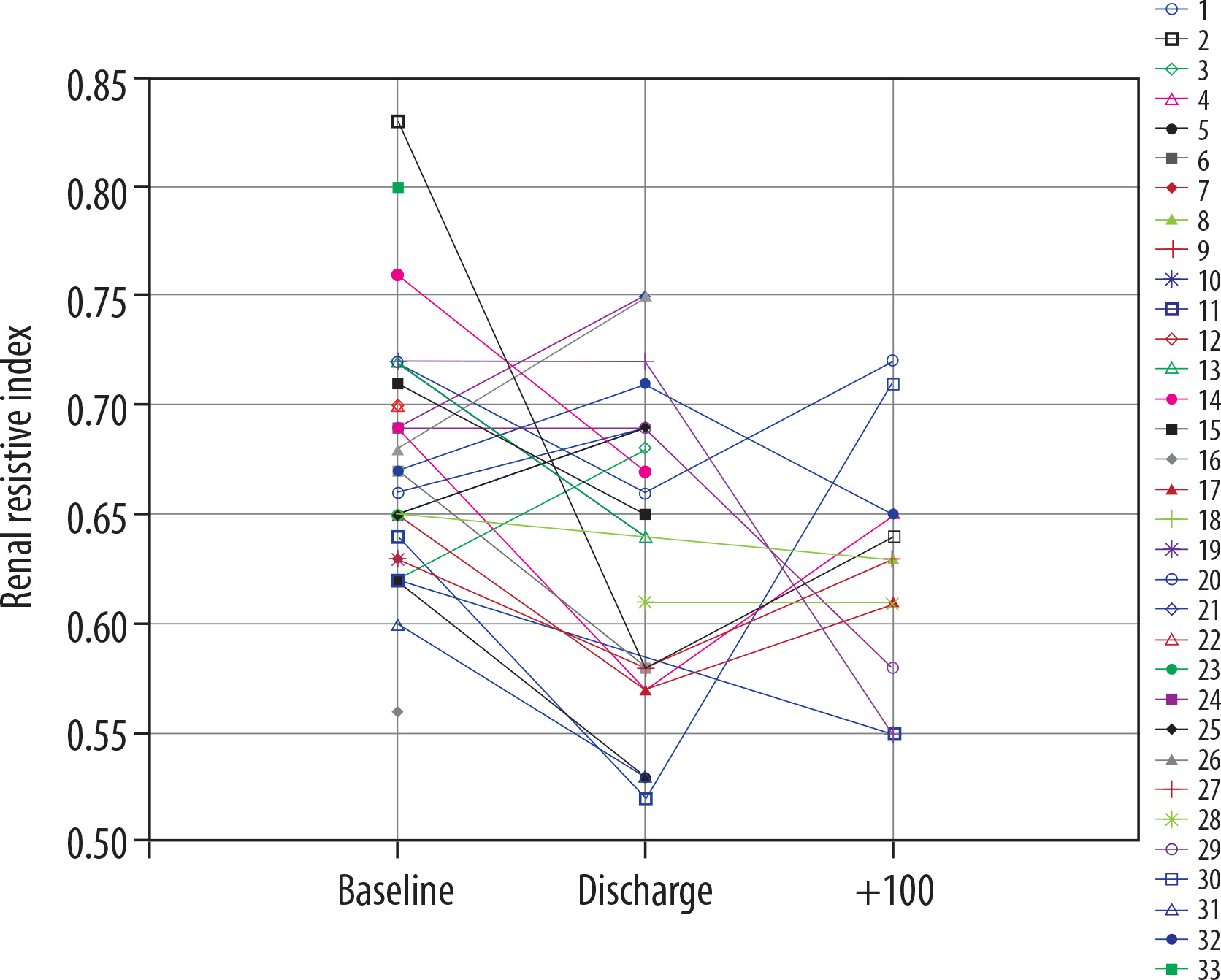
Figure 3 depicts case profiles of all measurements. We compared the results of the three successive measurements and found a statistically nonsignificant decrease of rRI (p = 0.12, Supplementary Figure 1).
Results of measurements and ratios of discharge/baseline and day +100/baseline were compared between patients who developed AKI, sepsis, and acute or chronic GvHD between baseline and the last measurement, but no significant differences were found (Supplementary Table 2). There was also no correlation of the measurements and their changes with CNI levels; however, the number of observations was relatively low: 23 for CSA and six for TAC (Supplementary Table 3).
rSWE
Case profiles of rSWE are shown in Figure 4. The values of the three successive measurements did not differ significantly (p = 0.77, Supplementary Figure 2).
Figure 4
Case profiles of renal shear wave elastography at three timepoints in all patients (A), patients treated with vasopressors (B), and patients with chronic graft versus host disease (cGvHD) after day +100 (C)
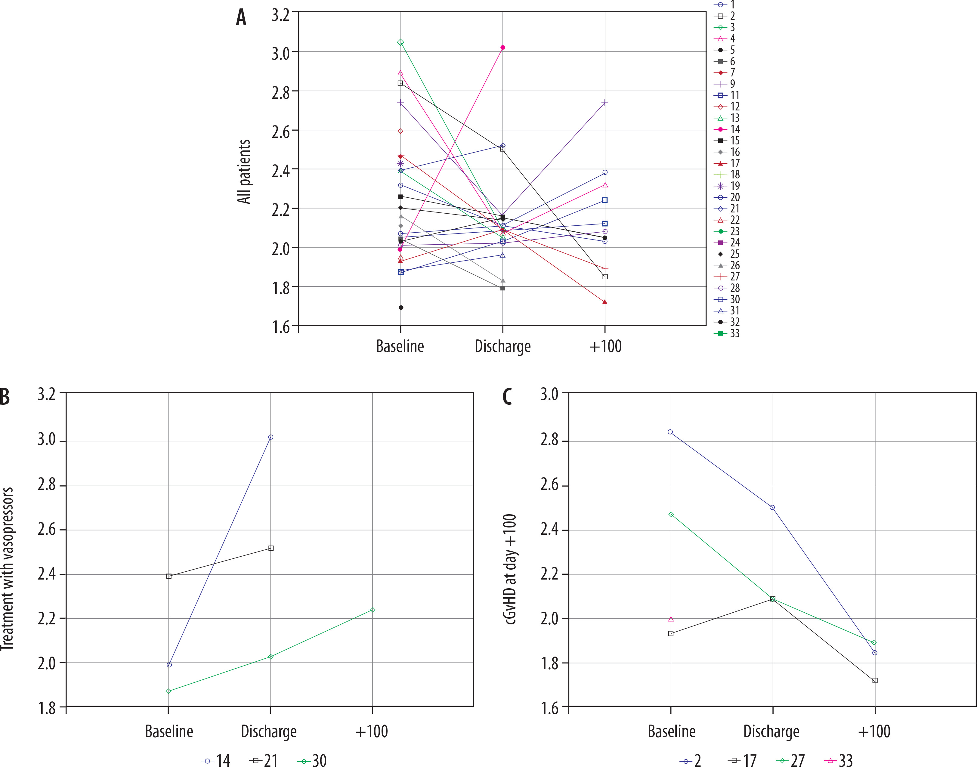
A case-by-case analysis revealed a marked increase of kidney elastography in three patients with serious inflammatory complications leading to hypotension requiring vasopressor treatment: patient 14 with anaphylactic shock secondary to platelet transfusion four days after alloHCT, patient 21 with septic shock at day 11, and patient 30 with grade three cytokine release syndrome in haploidentical donor transplantation and acute GvHD grade two diagnosed at the 18th day. Interestingly, none of these subjects met criteria for AKI but all had a significant decrease in eGFR after 100 days, compared to baseline (26.6%, 40.3%, 43.8%). The ratio of baseline/discharge rSWE was significantly higher in patients treated with vasopressors than the remaining group (p = 0.008, Figure 4B, Supplementary Figure 2). Since these were unique complications in the study population, these three cases were excluded from further analysis.
When the absolute values and ratios of repeated measurements to baseline were compared stratified by AKI, sepsis before day +100, and acute and chronic GvHD, we observed significantly lower rSWE +100 and rSWE +100/baseline ratio in patients with chronic GVHD (p = 0.012 and 0.024, respectively; median difference of rSWE +100 was 33%, 95% CI: 10-65%) (Figure 4C). However, this concerned only 3 out of 12 patients who had day +100 assessment.
Discussion
Findings of this study may be summarized as follows: 1) renal RI and rSWE were higher in patients scheduled for alloHCT than in the control group; 2) baseline rRI in alloHCT recipients correlated negatively with eGFR beyond 100 days after HCT; 3) renal SWE was significantly elevated in cases complicated with hypotension requiring vasopressor treatment; 4) renal SWE after day +100 was significantly lower in patients with chronic GvHD.
According to previous studies, factors associated with increasing rRI are: higher age, lower eGFR, diabetes, cardiovascular disorders, smoking, and CNI, while drugs inhibiting the renin-angiotensin-aldosterone system are associated with reduction of rRI [22,23]. The potential of CNI to increase rRI is reversible either by cessation of CNI or administration of nitroglycerin [24,25]. A decrease in rRI may also be a result of vasodilation secondary to inflammation that does not omit renal vessels [26]. Our findings did not align with all the above observations, as rRI did not correlate with CNI concentration, and it did not increase after introduction of CNI. This may be explained by extreme factors influencing alloHCT recipients: inflammatory process generated by the recovering transplanted immune system, severe infections, and fluctuating dosing of CNI depending on the risk of relapse, GvHD, infection, and time from transplantation. However, the correlation of baseline rRI with day +100 eGFR places rRI as a potential predictor of kidney dysfunction after alloHCT.
While rRI is a standard test performed in clinical practice, rSWE is currently used only in research. What is more, findings are diverse and in some cases contradictory. Numerous studies have found a negative correlation of rSWE and eGFR in the transplanted kidney and a positive correlation with extent of fibrosis assessed by renal biopsy. However, results in native kidneys are highly controversial in CKD: some indicate lower rSWE while others report increased rSWE. The results of a recent meta-analysis showed a non-significant difference in rSWE of –0.82 m/s in patients with CKD versus non-CKD patients [12]. This may be explained by heterogenous histological and hemodynamic changes secondary to underlying and concomitant diseases, medications, different techniques of measurement, and other factors that are poorly described. Although our study did not find correlations of rSWE and eGFR, it shed light on the possible influence of chronic GvHD that is independent of CNI concentration. The lower rSWE observed in chronic GvHD may potentially be explained by histological changes in the kidney induced by GvHD, postulated in numerous former studies [4,27-30].
Hypothetically, the reversible reduction of kidney elasticity may be due to the influence of CNI on vasoconstriction of afferent arterioles and consequent reduction of renal blood flow [25]. Our findings do not support this explanation, since CNI levels did not correlate with rSWE.
In the case of AKI, studies report that rSWE is higher in AKI as a result of renal parenchyma edema [31-33]. rSWE in our study did not differ in patients who experienced AKI or not, but the measurements were performed when patients were ambulatory and stable, not at the time of AKI. This suggests that AKI has no long-term influence on rSWE. However, it might be present in cases of severe hypotension treated with vasopressors, as observed in three patients from our study. Interestingly, none of these subjects met the criteria for AKI, but all experienced a significant decrease in eGFR after 100 days, compared to baseline. This suggests that acute hypoperfusion may have long-term consequences that do not manifest as AKI but are reflected in increased rSWE.
Our study has certain limitations; first of all, as a pilot study in a specific and heterogenous group with a high proportion of missing observations (only 13 out of 33 patients underwent 3 measurements), universal conclusions cannot be made. The number of missing observations is high because the measurements were performed in ambulatory patients with no uncontrolled medical conditions. This makes the observations less prone to the influence of acute conditions. However, we were not able to control for all previously reported confounding factors. The above-mentioned attrition bias together with lack of adjustment for comorbidities and concomitant treatments contributes to the limited statistical power of the study. Larger, adequately powered prospective studies are warranted to clarify our exploratory findings.
Secondly, unlike numerous similar studies, the intra- and inter-operator variability was not assessed, which is due to the vulnerable population of patients with impaired immunity requiring numerous assessments within the time of alloHCT.
The strength of the study is that measurements were performed at three time points, which allows analysis of changes across time. Moreover, all records were collected manually; therefore, reliable case-by-case analysis of this heterogenous group enabled accurate analysis of potential correlations that highlight areas for further research.
Conclusions
Baseline rRI and rSWE differed between patients scheduled for alloHCT and the control group, and both parameters fluctuated over time. Renal SWE is potentially influenced by severe hypotension and chronic GvHD. Further, larger-scale research on factors influencing the tests may be crucial to accurately explore the diagnostic potential of both methods.

