

Introduction
Since the publication of several randomized controlled trials (RCTs) in 2015, endovascular therapy (EVT) has become the standard of care in patients with acute stroke due to intracranial large vessel occlusions of the anterior circulation [1].
Internal carotid artery (ICA) occlusions account for 6-28% of all ischemic strokes [2-4], with occlusion of the carotid-T, i.e. the terminal part including the origins of the middle and anterior cerebral arteries, being a severe manifestation of the disease, as collateral flow to the ischemic territory is particularly compromised. Isolated, more proximal occlusions of the ICA without involvement of the carotid-T (non-terminal ICA [non-T ICA] occlusions) allow for various forms of collateralization, resulting in a more variable spectrum of stroke severity, ranging from mild to severe. The significance of collateral circulation in the context of acute stroke has been widely explored in previous research [5-8]. Notably, patients with large-artery atherosclerotic occlusions of the ICA tend to exhibit more robust collateral networks, likely as a result of chronic cerebral hypoperfusion, which facilitates both the development of new collaterals and the recruitment of pre-existing ones [9]. This compensatory mechanism contributes to the sustained viability of ischemic brain tissue, potentially leading to improved functional outcomes [10-13]. Nevertheless, the specific role of collaterals in non-T ICA occlusions remains insufficiently addressed in the current body of literature. Overall, acute ICA occlusions can be associated with poor prognosis, with the 90-day mortality reaching up to 22% [14,15]. Non-T ICA occlusions were not well represented or were absent in the aforementioned pivotal RCTs on endovascular stroke therapy. Retrospective series or registry data have yielded somewhat controversial results regarding this specific situation [16-23]. To date, no study has systematically analyzed the impact of intracranial collateral status and distal embolization on outcomes in non-T ICA occlusions undergoing EVT. Therefore, we sought to evaluate our experience with EVT for isolated non-T ICA occlusions from a tertiary referral stroke center regarding safety and efficacy and explore the role of collaterals and distal emboli in this specific occlusion pattern.
Material and methods
Patients and imaging
From a prospectively maintained institutional database, part of the multicenter German Stroke Registry for Endovascular Therapy (GSR-ET) [24], all patients treated with EVT for acute isolated non-T ICA occlusion between August 2015 and February 2023 were identified. Acute non-terminal ICA occlusions were diagnosed by computed tomography (CT) angiography or magnetic resonance angiography. EVT was pursued in patients with clinically relevant neurological deficits not explained by established focal infarctions on imaging or signs of hemodynamic instability (such as fluctuating or blood pressure-dependent deficits). Low Alberta Stroke Program Early CT Score (ASPECTS) and extended time window (> 24 h) were considered relative contraindications and were assessed on a case-by-case basis, as were mismatch profiles on CT perfusion indicating salvageable tissue at risk.
Patients with ICA terminus occlusion or any additional intracranial cerebral artery occlusions (tandem occlusions) were excluded from this study.
Clinical and demographic data were collected, including age, sex, cardiovascular stroke risk factors, and neurological and functional status (National Institutes of Health Stroke Scale [NIHSS] and modified Rankin Scale [mRS]) at admission and discharge. Good clinical outcome was defined as mRS ≤ 2, fair clinical outcome as mRS ≤ 3. Intracranial bleeding was characterized in accordance with the European Cooperative Acute Stroke Study III classification system. An early neurological improvement was defined as a decrease of 4 or more points on the NIHSS compared to the baseline. Conversely, early neurological deterioration was defined as an increase of 4 or more points on the NIHSS compared to the baseline.
Imaging data collection included ASPECTS at baseline and discharge and assessment of leptomeningeal collateral status on preinterventional CT angiography (CTA) according to the modified Tan (mTan) 4-point intracranial collateral score as described previously [25,26]: 0 (absent): absent collateral supply of the affected territory; 1 (poor): collateral supply filling ≤ 50%; 2 (moderate): collateral supply filling > 50% but < 100%; and 3 (good): 100% collateral supply of the occluded territory. The mTan score was assessed by one non-blinded, board-certified radiologist with 2 years of experience in neuroradiology.
Furthermore, data on occlusion site (extracranial, intracranial, or combined), etiology (atherosclerotic, cardioembolic, or dissection), and occlusion length were collected. Occlusions were dichotomized into short (partial cervical occlusion or involvement of a single intracranial ICA segment) vs. long (involvement of the entire cervical ICA segment, > 1 intracranial ICA segments, or combined extra-intracranial) segment occlusions. Recorded procedural details were EVT devices used, techniques applied, number of passes, angiographic outcome, time metrics, complications, and type of anesthesia.
The study was approved by the Charité Ethics Committee (EA2/082/15). Informed consent of individual patients or of the next of kin for the dead patients was waived for this retrospective study of anonymized data according to pertinent institutional guidelines.
The statistical analysis was performed using SPSS Statistics Version 26 for Windows (IBM, Armonk, New York, USA). The mean, median, standard deviation and interquartile ranges (IQR) were determined when appropriate. Statistical significance was calculated using χ2 tests and the Mann-Whitney U test. The tests were performed for the entire patient cohort as well as for subgroups stratified by the mTan score (mTan 1-2 vs. mTan 3). Given the low sample size and contingent risk for type-II error, p < 0.1 was considered indicative of a potential association in this exploratory analysis. Given the small sample size, we did not calculate adjusted odds ratios (ORs) to avoid model overfitting. However, given the equal distribution of age, pre-stroke disability and NIHSS as the most important outcome predictors, unadjusted ORs may provide an approximation of the association between collateral status and clinical outcomes or complications such as intracranial hemorrhage (ICH).
Endovascular procedure
Procedures were performed by a team of 7 neurointerventionalists. Procedural details varied depending on occlusion site and type, as well as operator preference. Treatments were performed under conscious sedation or general anesthesia. In general, all interventions were performed via transfemoral access with 8 or 9F sheaths. Balloon guide catheters (Cello or FlowGate2), distal aspiration catheters (SOFIA 5 and 6F) and a variety of stent retrievers (Solitaire 6x40, Preset 6x30, EmboTrap II) were used in most of the cases. Stent angioplasty was performed in cases of identifiable high-grade, ulcerated, or re-occluding stenosis, or in cases of signs of hemodynamic compromise (especially in dissecting stenoses, after assessment of contralateral and posterior circulation collaterals) either extra- or intracranially following EVT (extracranial stent types included Carotid WALLSTENT, C-Guard and Leo; intracranial stent types included Accero, Credo Stents, and Leo). The choice of stent as well as antiplatelet medication (mostly 250-500 mg aspirin or a half-dose, body-weight-adjusted bolus regimen of Tirofiban) was at the neurointerventionalists’ discretion taking both estimated infarct size, premedication, and systemic thrombolysis status into consideration. In cases of distal embolization, additional EVT was pursued if deemed both feasible and reasonably safe.
Results
Patients and imaging
Results are summarized in Table 1. The study cohort consisted of 58 patients treated for acute non-T ICA occlusions. The median NIHSS at admission was 13 (IQR: 7-18). Twenty-five patients (43.1%) received bridging intravenous thrombolysis therapy prior to EVT. Twenty patients (39.2 %) presented with a good modified intracranial collateral score (mTan 3) on CTA, whereas 22 patients (43.1%) had moderate (mTan 2) and 9 patients had poor (mTan 1) collaterals (17.6%).
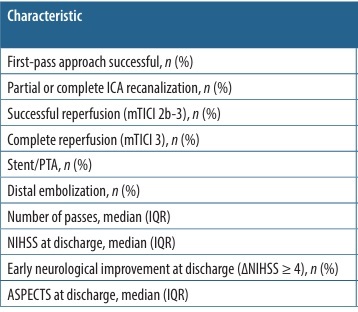
Table 1
Demographic, clinical, imaging, procedural, and outcome characteristics
[i] ASPECTS – Alberta Stroke Program Early CT Score, ICA – internal carotid artery, IQR – interquartile range, i.v. – intravenous, mTan – modified Tan intracranial collateral score, NIHSS – National Institutes of Health Stroke Scale, PTA – percutaneous transluminal angioplasty, SD – standard deviation
Stroke etiology was cardioembolism, large-artery atherosclerosis, and dissection in 30 (51.7%), 21 (36.2 %), and 6 (10.4%) patients, respectively. One patient (1.7%) had sarcoma infiltration of the petrous canal. Approximately half of the patient population (30, 51.7%) presented with an intracranial ICA occlusion and 15 patients (25.8%) with an isolated extracranial ICA occlusion. Combined occlusions were found in 13 patients (22.4%).
Most patients showed no or only minimal early ischemic changes on the initial scan, with a median ASPECTS of 10 (IQR: 8-10).
EVT procedures and angiographic outcomes
Most patients (45, 77.6%) were treated under general anesthesia, while the remaining patients underwent conscious sedation.
Proximal flow control using a balloon guide catheter was performed in 77.6% of cases. Angioplasty (balloon angioplasty, percutaneous transluminal angioplasty, or stenting) of the ICA following thrombectomy was performed in 22 patients (37.9%), including 3 of 6 patients with dissection (50%) and 16 of 21 patients with atherosclerotic occlusions. A first-pass approach achieved successful reperfusion in 26 patients (44.8%). The median number of passes was 2 (IQR: 1-3). Successful ICA reperfusion (modified Thrombolysis in Cerebral Infarction [mTICI] 2b-3) was achieved in 53 patients (91.4%).
Clinical and imaging outcome
The median ASPECTS on follow-up imaging was 8 (IQR: 6-9). Two patients developed posterior cerebral artery (PCA) infarction due to embolization via the posterior communicating artery (PComA) and two patients developed an anterior cerebral artery (ACA) infarction (one of these despite angiographically successful rescue EVT and one with a distal embolization not rescued by EVT).
Median NIHSS 24 h after the EVT and at discharge was 10 (IQR: 6-21) and 3 (IQR: 2-9), respectively. At discharge, early neurological improvement was found in 7 (53.8%) patients with a poor to medium mTan score and in 8 patients (66.7%) with a good collateral score (p = 0.668). No cases of early neurological deterioration at discharge were identified.
The in-hospital mortality of the cohort was 20.7% (12 patients). After excluding patients with pre-stroke disability (mRS > 2), a good clinical outcome (mRS ≤ 2) on discharge and at 90-day follow-up was achieved in 17 (41.5%) and 10 (41.7%) patients, respectively.
Safety aspects
In 13 (22.4%) patients, thrombus migration or distal embolism occurred during the procedure, including 6 intracranial non-T occlusions, 6 isolated extracranial ICA non-T occlusions and in 1 combined occlusion. Distal embolism affected the M1 segment in 2 patients, the M2 segment in 5 patients, the A2 segment in 2 patients, and combined ACA/middle cerebral artery (MCA) branch occlusions in 2 patients, as well as the PCA via the PComA in 2 patients. Nine of these 13 patients underwent additional EVT for distal embolization, with 5 patients (38.5%) achieving mTICI 3 reperfusion and 8 (61.5%) achieving mTICI 2b-3 reperfusion. No complications occurred in rescue EVT maneuvers for procedural distal embolism. In 4 patients, distal rescue EVT was not performed because it was not deemed safe or feasible by the interventionalist. The median time from distal embolization to distal reperfusion was 19 min (IQR: 10-26).
ICH occurred in 10 patients (17.2%). One remote hemorrhage was attributed to intravenous thrombolysis. Three patients (5.2%) developed a symptomatic ICH (sICH), 2 of which required a decompressive hemicraniectomy. Non-flow limiting ICA dissections occurred in 5 patients (8.6%) requiring medical therapy (dual antiplatelet medication) only. One iatrogenic dissection was treated with a stent due to relevant flow limitation.
Impact of collateral scores
Results are summarized in Tables 2 and 3. Notably, patients with good collateral scores showed a potential association with higher ASPECTS at admission compared to those with moderate to poor collateral status (10 [IQR: 9-10] vs. 9 [IQR: 7-10], p = 0.086). At discharge, patients with good collateral status had significantly higher ASPECTS compared to patients with poor to moderate collateral status (9 [IQR: 6-10] vs. 7 [IQR: 5-9], p = 0.035).
Table 2
Procedural characteristics, imaging findings, and clinical outcomes, according to modified Tan intracranial collateral (mTan) score
Table 3
Clinical outcomes at discharge and 90-day follow-up according to modified Tan intracranial collateral (mTan) score
There was a potential association between good collateral status and a higher proportion of patients achieving mRS ≤ 3 at discharge in the good collateral score group (55.6% vs. 31%; OR 2.78; 95% confidence interval [CI]: 0.82-9.39; p = 0.10). Further results for clinical outcome showed a similar pattern but did not reach statistical significance. There was a numerically higher rate of good clinical outcome (mRS ≤ 2) at discharge in patients with a good collateral status (38.9% vs. 27.6%, OR 1.67 [95% CI: 0.48-5.83], p = 0.42). At three-month follow-up, patients with good and poor to moderate collateral score presented with mRS ≤ 2 in 45.5% and 26.3%, respectively (OR 2.33; 95% CI: 0.49-11.17; p = 0.29) and with mRS ≤ 3 in 54.5% and 31.6%, respectively (OR 2.6; 95% CI: 0.56-12.02; p = 0.22). Patients with good collateral status had a numerical lower mortality at discharge (16.7% vs. 27.6%, OR 0.53; 95% CI: 0.12-2.31; p = 0.39) and at three months (27.3% vs. 52.6%, OR 0.34; 95% CI: 0.97-1.68; p = 0.18), but this observation was not significant.
There was no association between stroke etiology and the intracranial collateral score. Patients with good collateral status had lower rates of any ICH (15.8% vs. 23.3%, p = 0.53) and sICH (5.3% vs. 10% 0.56); however, these differences did not reach statistical significance.
Impact of distal embolization
Results are summarized in Table 4. Patients with complete reperfusion of distal emboli (mTICI 3) demonstrated significantly better functional outcomes at discharge and at 90 days compared to those with incomplete distal reperfusion (mTICI 2b/c). At discharge, 100% of patients with complete distal reperfusion (mTICI 3) achieved mRS ≤ 2 and mRS ≤ 3 compared to 0% and 50% (p = 0.008 and 0.025), respectively, in patients with incomplete reperfusion (mTICI 2b/c). Similarly, at 90 days, 100% of patients with complete reperfusion of distal emboli showed mRS ≤ 2 and mRS ≤ 3 compared to 0% and 0% (p = 0.028 and 0.049), respectively, for those with incomplete reperfusion of distal emboli (mTICI 2b/c). There was a potential association between distal embolization and higher NIHSS scores at discharge in patients with distal embolization compared to patients without distal embolization or those with complete reperfusion (p = 0.094). No significant differences were observed in rates of early neurological improvement, ASPECTS at discharge, mortality, or hemorrhage.
Table 4
Imaging and clinical outcomes according to distal embolization and subsequent reperfusion status
[i] ASPECTS – Alberta Stroke Program Early CT Score, ECASS III – European Cooperative Acute Stroke Study III, ICH – intracranial hemorrhage, IQR – interquartile range, mRS – modified Rankin Scale, mTICI – modified Thrombolysis in Cerebral Infarction, NIHSS – National Institutes of Health Stroke Scale, sICH – symptomatic ICH
Discussion
In this retrospective observational study, we describe our experience at a high-volume tertiary care referral center with EVT of acute symptomatic non-T ICA occlusions in 58 consecutive patients. We show that EVT of non-T ICA occlusions is technically effective, with a high rate of technical success (successful reperfusion [mTICI 2b-3] in 91% of patients) and a good procedural safety profile (sICH rate of 5%). Reservations regarding EVT in non-T occlusion may in part result from consideration of causing distal emboli with flow obstruction distal to the Circle of Willis thereby leading to aggravation of symptoms. While such emboli occurred in a non-negligible proportion of cases (22%) despite the use of proximal flow control, the majority of these cases (62%) could be retrieved. Our data suggest that complete distal reperfusion (mTICI 3) may be associated with better functional outcomes compared to incomplete reperfusion (mTICI 2b/c); however, these findings are based on small subgroup analyses and should be interpreted cautiously. This observation may indicate that achieving complete distal reperfusion could be beneficial, although this hypothesis requires confirmation in larger prospective cohorts. Although no significant differences were observed in mortality or hemorrhage rates, the potential association between complete distal reperfusion and lower NIHSS scores may indicate better neurological recovery. These findings emphasize the need for strategies focused on minimizing and fully retrieving distal emboli to improve clinical outcomes. In contrast to M1 occlusions, where mTICI 2b/2c may already represent a clinically meaningful result, in non-T ICA occlusions with open distal intracranial vessels, only complete perfusion (mTICI 3) may be considered a sufficient angiographic endpoint. Accordingly, attempting thrombectomy of distal emboli arising during mechanical thrombectomy of the distal ICA may be reasonable, although this observation requires further validation.
Although the procedural results were encouraging, the clinical outcomes were more modest. At the 90-day follow-up, only 42% of patients achieved a good outcome (mRS ≤ 2), and the in-hospital mortality rate was 21%. These results are comparable with the outcomes presented in several studies, including GSR-ET and International Registry of Endovascular Treatment in Acute Stroke (IRETAS), which also showed rather low rates of good clinical outcome after 3 months (28-56%), in-hospital mortality ranging from 24% to 29%, and sICH in 3-9% [21,27,28]. Furthermore, our study highlights a potential role of collateral status in thrombectomy of non-T-ICA occlusions, with higher ASPECTS (9 vs. 7) and a higher percentage of patients with mRS ≤ 2 at discharge (39% vs. 28%) and at 90 days (46% vs. 26%) in patients with good collaterals compared to those with poor to moderate collateral status.
So far, there are no RCTs concerning EVT for acute non-T ICA occlusions, and the number of such occlusions included in previous RCTs is negligible [29-33]; therefore, the available evidence remains limited and is derived mainly from retrospective studies and case series. The results of our study concerning the angiographic outcomes, functional outcomes, and safety are similar to those reported in previous literature. Reported rates of successful reperfusion for ICA occlusions treated with EVT range from 68.7% to 92% [19-21,34,35]. Rates were reported to be lower for extracranial (69%-77%) [20,21,35] and isolated intracranial ICA occlusions (56%-80%) [18,21]. Our distal embolization rate of 21% aligns with previous reports of 20-22% [18,34] but is higher than the Intravenous thrombolysis or endovascular treatment for acute ischemic stroke associated with cervical internal carotid artery occlusion (ICARO-3) and German Stroke Registry rates [21]. In-hospital mortality (21%) and good functional outcomes (mRS < 2) in our cohort (42%) are within the expected range but lower compared to some studies with younger patients [34]. Our findings align with the multicenter Acute Carotid Occlusion Below Circle of Willis (ACOBOW) study by Meyer et al. [23], which also reported high technical success rates but modest functional outcomes in non-T ICA occlusions. Unlike ACOBOW, which compared EVT with best medical treatment and found no significant overall superiority, our study provides more granular insights into the prognostic role of collateral circulation and the importance of complete rescue of distal emboli.
In contrast to the Endovascular Treatment for Isolated Internal Carotid Artery Occlusion (ETIICA) and ACOBOW studies, which primarily assessed clinical efficacy of EVT versus medical management, our analysis provides mechanistic insight into factors determining outcome in non-T ICA occlusions. Specifically, we demonstrate that collateral status strongly influences imaging preservation and that only complete reperfusion (mTICI 3) after distal embolization translates into favorable recovery. These findings refine procedural understanding by suggesting that technical success in non-T ICA occlusions requires not merely vessel reopening but full downstream reperfusion, a nuance not captured in previous multicenter reports.
The mTan collateral score has shown value in the assessment of collateral status in patients suffering from proximal MCA occlusion. However, to our knowledge, this is the first study to evaluate the mTan collateral score specifically in patients with non-T-ICA occlusions. In this situation, collaterals are of pivotal importance and include not only pial collaterals but potentially also collaterals via the anterior communicating artery, PComA, or ophthalmic artery depending on the occlusion site and individual vascular anatomy. Our study highlights the critical role of collateral circulation in patients with non-T ICA occlusions undergoing EVT. It demonstrates that those with higher mTan collateral scores exhibit improved imaging outcomes (ASPECTS at discharge) and suggests better clinical results at both discharge and the 90-day follow-up, alongside reduced mortality. This finding persists even in patients presenting with clinically significant deficits, despite having favorable ASPECTS and mTan scores. The importance of collateral circulation to ensure perfusion of the ischemic tissue in cases of large vessel occlusion and its prognostic value has already been extensively discussed in the past [5-8]; however, to our knowledge, it has not yet been specifically investigated for non-T ICA occlusions. Three primary factors may influence a non-T ICA occlusion patient’s neurological status upon admission. First, small distal emboli, which are often undetectable on CTA, may contribute to neurological impairment, while their respective infarcts may be missed on CT. In this situation, effective collateral circulation may play a critical role in maintaining perfusion and limiting infarct burden. Second, systemic blood pressure and hemodynamic insufficiency compromise cerebral perfusion, making the status of intracranial collaterals crucial for maintaining cerebral perfusion. Third, a combination of distal embolization and hemodynamic compromise may contribute to clinical presentation. In clinical practice, the decision-making process in non-T ICA occlusion treatment remains challenging and is less straightforward as compared to other occlusion locations, especially if the patient is only moderately affected. The risks of neurological worsening due to loss of collateral support, especially in asymptomatic or low-symptomatic patients, should be outweighed by the potential hazards of EVT treatment, especially distal embolization. In this specific situation, achieving complete reperfusion (mTICI 3) may be particularly important given the presence of otherwise patent distal intracranial vessels.
The observed 100% favorable outcome rate in patients with successfully rescued distal embolization must be interpreted with extreme caution due to the very small subgroup size and potential selection effects. This finding should be regarded as hypothesis-generating only and does not imply a causal benefit of procedural embolization or rescue therapy. This apparent discrepancy should therefore be interpreted with caution and cannot be taken as evidence that procedural embolization itself is beneficial. Rather, it underlines the variability inherent in small subgroups and highlights that complete distal reperfusion (mTICI 3) can normalize outcomes when distal emboli occur, whereas incomplete rescue may contribute to poor recovery.
Our study shows a low rate of periprocedural complications, suggesting that EVT in non-T ICA occlusions may be a reasonably safe and technically effective procedure. While the annual risk of ipsilateral stroke in persistent ICA occlusion has been reported to be substantial (7.7%) [36], this figure reflects long-term risk of recurrent stroke rather than evidence for acute interventional intensification. Therefore, the potential role of more aggressive acute treatment strategies in non-T ICA occlusions should be considered exploratory and warrants further prospective investigation. Although patients with poor to moderate collateral status exhibited less favorable imaging and clinical outcomes, a lower mTan score should not preclude the use of EVT in non-T ICA occlusions. The observed differences in outcomes were relatively subtle, and the potential for significant worsening without interventional therapy may be non-negligible. Another critical aspect is careful blood pressure management in patients with poor collateral circulation. Avoiding hypotension and maintaining adequate blood pressure may be important to preserve cerebral perfusion and limit further ischemic injury.
The novelty of our study lies in two main aspects. First, to our knowledge, this is among the first studies to systematically evaluate intracranial collateral status using the m-Tan score specifically in non-T ICA occlusions, suggesting that it may have prognostic relevance for both imaging and clinical outcomes in this unique subgroup. Second, we highlight the critical importance of distal embolization management: while distal emboli occurred in a relevant proportion of cases, patients in whom complete distal reperfusion (mTICI 3) was achieved after rescue EVT showed markedly better functional outcomes than those with incomplete reperfusion. Together, these findings emphasize that EVT in non-T ICA occlusions should aim not only at restoring ICA patency, but also at securing full downstream reperfusion, with collateral assessment serving as a key element in treatment selection and periprocedural strategy.
The main limitations of this study are its retrospective monocentric character with its inherent limitations, the moderate sample size increasing the risk of type-2 error, selection bias towards more severely affected patients, and a lack of a control group being treated with best medical therapy for comparison. We thus cannot infer from our data whether non-T ICA occlusions should undergo EVT in general or not. The limited sample size prevented multivariate adjustment and may have led to random fluctuations in subgroup outcomes. Several subgroup analyses, particularly regarding distal embolization management and mTICI subcategories, are based on small patient numbers and should therefore be considered exploratory and hypothesis-generating. Furthermore, the mTan score was assessed by only one non-blinded reader.
Conclusions
Our results suggest that EVT in isolated non-T ICA occlusions is technically feasible and can achieve high reperfusion rates with an acceptable safety profile in selected patients, although functional outcomes remain variable. Given the retrospective design and absence of a control group, conclusions regarding comparative effectiveness must be drawn cautiously. Furthermore, our findings suggest an association between good collateral status and improved imaging and the clinical outcomes in patients with non-T-ICA occlusions. However, the comparative clinical effectiveness of EVT versus best medical management remains to be determined in further RCTs.

