

GASTROINTESTINAL AND ABDOMINAL RADIOLOGY / ORIGINAL PAPER
Factors associated with diaphragmatic invasion in hepatic alveolar echinococcosis: a retrospective CT-based analysis
1
Radiology Department, I.K. Akhunbaev Kyrgyz State Medical Academy, Bishkek, Kyrgyz Republic
2
Department of Diagnostic and Interventional Radiology, Caritas Klinikum Saarbrücken, Academic Teaching Hospital of Saarland University, Saarbrücken, Germany
Submission date: 2025-12-18
Final revision date: 2026-04-06
Acceptance date: 2026-04-18
Publication date: 2026-10-23
Corresponding author
Iliar Baudinov
Department of Radiology and Therapy I.K. Akhunbaev Kyrgyz State Medical Academy 92 Akhunbaev Street, Bishkek, Kyrgyz Republic
Department of Radiology and Therapy I.K. Akhunbaev Kyrgyz State Medical Academy 92 Akhunbaev Street, Bishkek, Kyrgyz Republic
Pol J Radiol, 2026; 91(1): 506-513
KEYWORDS
risk factorsdiaphragmmultidetector computed tomographyhepatic alveolar echinococcosissurgical planningorgan invasion
TOPICS
ABSTRACT
Purpose:
The purpose of this study was to determine the frequency of diaphragmatic invasion and to identify topographic, clinical, and morphological factors associated with it using contrast-enhanced computed tomography (CT).
Material and methods:
In this retrospective study, 201 patients with confirmed hepatic alveolar echinococcosis (AE) examined at national referral centres between 2018 and 2025 were included. All patients underwent multiphase contrast-enhanced multidetector CT of the liver. Demographic data, lesion number and localisation, Alveolar Echinococcosis Ulm Classification type, maximum lesion diameter, vascular invasion, and extrahepatic extension were recorded. Diaphragmatic invasion was defined by the presence of at least two predefined CT criteria. Associations were assessed using Cramer’s V, and predictors of diaphragmatic invasion were analysed using multivariable logistic regression.
Results:
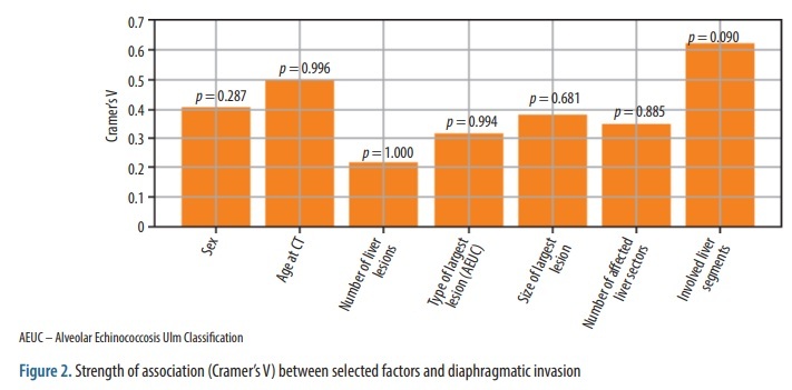
Extrahepatic spread was identified in 108 patients (53.7%). The association between diaphragmatic invasion and involved liver segments did not reach statistical significance (Cramer’s V = 0.619; p = 0.090). This result should be interpreted cautiously. Diaphragmatic involvement was more frequently observed in patients with lesions affecting the superior liver segments S2, S7, and S8. No significant positive associations were found between diaphragmatic invasion and vascular involvement (Cramer’s V ≤ 0.484; all p > 0.65). In multivariable analysis, portal vein trunk invasion was inversely associated with diaphragmatic involvement (OR = 0.234; 95% CI: 0.062-0.883; p = 0.032).
Conclusions:
Diaphragmatic invasion is a relatively frequent feature of hepatic AE. Associations with liver segment involvement and vascular invasion require cautious interpretation. Recognition of characteristic CT findings remains important for pre-operative assessment and surgical planning.
The purpose of this study was to determine the frequency of diaphragmatic invasion and to identify topographic, clinical, and morphological factors associated with it using contrast-enhanced computed tomography (CT).
Material and methods:
In this retrospective study, 201 patients with confirmed hepatic alveolar echinococcosis (AE) examined at national referral centres between 2018 and 2025 were included. All patients underwent multiphase contrast-enhanced multidetector CT of the liver. Demographic data, lesion number and localisation, Alveolar Echinococcosis Ulm Classification type, maximum lesion diameter, vascular invasion, and extrahepatic extension were recorded. Diaphragmatic invasion was defined by the presence of at least two predefined CT criteria. Associations were assessed using Cramer’s V, and predictors of diaphragmatic invasion were analysed using multivariable logistic regression.
Results:
Extrahepatic spread was identified in 108 patients (53.7%). The association between diaphragmatic invasion and involved liver segments did not reach statistical significance (Cramer’s V = 0.619; p = 0.090). This result should be interpreted cautiously. Diaphragmatic involvement was more frequently observed in patients with lesions affecting the superior liver segments S2, S7, and S8. No significant positive associations were found between diaphragmatic invasion and vascular involvement (Cramer’s V ≤ 0.484; all p > 0.65). In multivariable analysis, portal vein trunk invasion was inversely associated with diaphragmatic involvement (OR = 0.234; 95% CI: 0.062-0.883; p = 0.032).
Conclusions:
Diaphragmatic invasion is a relatively frequent feature of hepatic AE. Associations with liver segment involvement and vascular invasion require cautious interpretation. Recognition of characteristic CT findings remains important for pre-operative assessment and surgical planning.
REFERENCES (12)
1.
Vuitton DA, McManus DP, Rogan MT, Romig T, Gottstein B, Naidich A, et al. International consensus on terminology to be used in the field of echinococcoses. Parasite 2020; 27: 41. DOI: 10.1051/parasite/2020024.
2.
Liu C, Fan H, Ge RL. A case of human hepatic alveolar echinococcosis accompanied by lung and brain metastases. Korean J Parasitol 2021; 59: 291-296.
3.
Casulli A, Abela B, Petrone D, et al. Unveiling the incidences and trends of alveolar echinococcosis in Europe: a systematic review from the KNOW-PATH project. Lancet Infect Dis 2026; 26: e49-e61. DOI: 10.1016/S1473-3099(25)00283-X.
4.
Gottstein B, Deplazes P. Alveolar echinococcosis: what triggers emergence in North America, Central Europe and Asia? Curr Opin Infect Dis 2021; 34: 440-446.
5.
Raimkulov K, Toigombaeva V, Kuttubaev O, Mergenov A. Assessment of echinococcosis invasion and behavioural risk factors among the population of Osh region of the Kyrgyz Republic. Euroasian Health J 2024; 16: 137-145. doi:10.54890/EHJ-2024-1-137.
6.
Paternoster G, Boo G, Wang C, Minbaeva G, Usubalieva J, Raimkulov KM, et al. Epidemic cystic and alveolar echinococcosis in Kyrgyzstan: an analysis of national surveillance data. Lancet Glob Health 2020; 8: e603-e611. DOI: 10.1016/S2214-109X(20)-30038-3.
7.
Wen H, Vuitton L, Tuxun T, Li J, Vuitton DA, Zhang W, McManus DP. Echinococcosis: advances in the 21st century. Clin Microbiol Rev 2019; 32: e00075-18. DOI: 10.1128/CMR.00075-18.
8.
Baudinov IA, Kadyrova AI, Omorov K, Bebezov BK, Ibraimov K, Oginskiy A, et al. Evaluation of vascular invasion in hepatic alveolar echinococcosis using computed tomography: an observational study. Heart Vessels Transplant 2025; 9. DOI: 10.24969/hvt.2024.553.
9.
Omorov RA, Aitbaev SA, Kanietov AK, Abdiev AA. Results of surgical treatment of patients with liver alveolar echinococcosis. Ann HPB Surg 2018; 23: 74-79.
10.
Bebezov BK, Bebezov KS, Umetaliev TM, Mamashev ND, Surov EA, Ryspekov BZ, Azhibekov SZ. Extended liver resections for alveococcosis. Vestn KRSU 2022; 22: 23-29.
11.
Brunetti E, Kern P, Vuitton DA. Expert consensus for the diagnosis and treatment of cystic and alveolar echinococcosis in humans. Acta Trop 2010; 114: 1-16.
12.
Graeter T, Schmidberger J. Stage-oriented CT classification and intermodal evolution model in hepatic alveolar echinococcosis. Rofo 2022; 194: 532-544.
Share
RELATED ARTICLE
| ISSN: | 1899-0967 |

We process personal data collected when visiting the website. The function of obtaining information about users and their behavior is carried out by voluntarily entered information in forms and saving cookies in end devices. Data, including cookies, are used to provide services, improve the user experience and to analyze the traffic in accordance with the Privacy policy. Data are also collected and processed by Google Analytics tool (more).
You can change cookies settings in your browser. Restricted use of cookies in the browser configuration may affect some functionalities of the website.
You can change cookies settings in your browser. Restricted use of cookies in the browser configuration may affect some functionalities of the website.