
GASTROINTESTINAL AND ABDOMINAL RADIOLOGY / ORIGINAL PAPER
Improved classification of benign and malignant hepatic tumors using combined ADC and T2-weighted radiomic features

1
Department of Radiology, University of Medicine and Pharmacy, Hue University, Hue, Vietnam
These authors had equal contribution to this work
Submission date: 2026-01-22
Final revision date: 2026-02-28
Acceptance date: 2026-03-15
Publication date: 2026-10-19
Corresponding author
Thanh Thao Nguyen
Department of Radiology, University of Medicine and Pharmacy, Hue University, Hue, Vietnam
Department of Radiology, University of Medicine and Pharmacy, Hue University, Hue, Vietnam
Pol J Radiol, 2026; 91(1): 495-505
KEYWORDS
TOPICS
ABSTRACT
Purpose:
To utilize machine learning algorithms to differentiate between malignant and benign hepatic lesions by integrating T2-weighted MRI-based radiomics and apparent diffusion coefficient (ADC) metrics.
Material and methods:
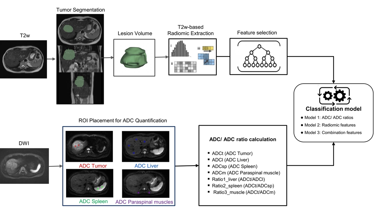
This cross-sectional study (January 2023–December 2024) enrolled 111 patients (71 male, 40 female) aged ≥ 18 years. Hepatic tumors were classified via radiological, pathological, and biochemical assessment. Radiomic features were derived from T2-weighted images, and ADC metrics from diffusion-weighted imaging. The classification performance was evaluated using three models: Model 1 (ADC and ADC Ratios), Model 2 (Radiomic Features), and Model 3 (Combined Features). Stratified 5-fold cross-validation was performed across four algorithms: Logistic Regression (LR), Support Vector Machine (SVM), Gradient Boosting (GB), and Random Forest. The area under the receiver operating characteristic curve (AUC) was used to measure the models’ discriminatory ability. All statistical analyses were conducted using Python libraries.
Results:
A total of 111 hepatic lesions were analyzed: 75 malignant and 36 benign. Among 12 significant radiomic features (p < 0.05), firstorder_InterquartileRange, ngtdm_Contrast, and glszm_ZonePercentage showed the largest effect sizes (Cohen’s d > 0.70). All ADC-related metrics were significantly higher in benign lesions compared to malignant ones (p < 0.001). Model 3 demonstrated the strongest overall performance, with both LR and SVM achieving accuracy and AUC values above 0.800. While Model 1 with SVM offered the highest sensitivity, Model 2 displayed the lowest specificity and weakest overall performance, whereas Model 3 generally showed higher classification performance than either feature set alone.
Conclusions:
Combining T2w-based radiomics features with ADC and ADC ratio values may improve discrimination between malignant and benign liver tumors.
To utilize machine learning algorithms to differentiate between malignant and benign hepatic lesions by integrating T2-weighted MRI-based radiomics and apparent diffusion coefficient (ADC) metrics.
Material and methods:
This cross-sectional study (January 2023–December 2024) enrolled 111 patients (71 male, 40 female) aged ≥ 18 years. Hepatic tumors were classified via radiological, pathological, and biochemical assessment. Radiomic features were derived from T2-weighted images, and ADC metrics from diffusion-weighted imaging. The classification performance was evaluated using three models: Model 1 (ADC and ADC Ratios), Model 2 (Radiomic Features), and Model 3 (Combined Features). Stratified 5-fold cross-validation was performed across four algorithms: Logistic Regression (LR), Support Vector Machine (SVM), Gradient Boosting (GB), and Random Forest. The area under the receiver operating characteristic curve (AUC) was used to measure the models’ discriminatory ability. All statistical analyses were conducted using Python libraries.
Results:
A total of 111 hepatic lesions were analyzed: 75 malignant and 36 benign. Among 12 significant radiomic features (p < 0.05), firstorder_InterquartileRange, ngtdm_Contrast, and glszm_ZonePercentage showed the largest effect sizes (Cohen’s d > 0.70). All ADC-related metrics were significantly higher in benign lesions compared to malignant ones (p < 0.001). Model 3 demonstrated the strongest overall performance, with both LR and SVM achieving accuracy and AUC values above 0.800. While Model 1 with SVM offered the highest sensitivity, Model 2 displayed the lowest specificity and weakest overall performance, whereas Model 3 generally showed higher classification performance than either feature set alone.
Conclusions:
Combining T2w-based radiomics features with ADC and ADC ratio values may improve discrimination between malignant and benign liver tumors.
REFERENCES (28)
1.
Salem S, Houseni M, Zidan L, Kandil A. The added value of PET/Ce-CT/DW-MRI fusion in assessment of hepatic focal lesions: PET/Ce-CT/DW-MRI fusion in hepatic focal lesion. Nucl Med Biol 2015; 42: 637-642.
2.
Sung H, Ferlay J, Siegel RL, Laversanne M, Soerjomataram I, Jemal A, et al. Global cancer statistics 2020: GLOBOCAN estimates of incidence and mortality worldwide for 36 cancers in 185 countries. CA Cancer J Clin 2021; 71: 209-249.
4.
Maino C, Vernuccio F, Cannella R, Cortese F, Franco PN, Gaetani C, et al. Liver metastases: the role of magnetic resonance imaging. World J Gastroenterol 2023; 29: 5180-5197.
5.
Bammer R. Basic principles of diffusion-weighted imaging. Eur J Radiol 2003; 45: 169-184.
6.
Koh DM, Collins DJ. Diffusion-weighted MRI in the body: applications and challenges in oncology. Am J Roentgenol 2007; 188: 1622-1635.
7.
Nalaini F, Shahbazi F, Mousavinezhad SM, Ansari A, Salehi M. Diagnostic accuracy of apparent diffusion coefficient (ADC) value in differentiating malignant from benign solid liver lesions: a systematic review and meta-analysis. Br J Radiol 2021; 94: 20210059. DOI: 10.1259/bjr.20210059.
8.
Sharma S, Dalal V, Prem Kumar G, Malhotra P. Diffusion MRI with quantification of ADC value in characterization of benign and malignant hepatic lesions and their correlation with cyto-histopathology. IP Int J Med Paediatr Oncol 2020; 6: 131-135.
9.
Mayerhoefer ME, Materka A, Langs G, Häggström I, Szczypiński P, Gibbs P, et al. Introduction to radiomics. J Nucl Med 2020; 61: 488-495.
10.
Baishya NK, Baishya K, Baishya K, Sarma R, Ray S. MRI radiomics in imaging of focal hepatic lesions: a narrative review. Cureus 2024; 16: e62570. DOI: 10.7759/cureus.62570.
11.
Starmans MPA, Miclea RL, Vilgrain V, Ronot M, Purcell Y, Verbeek J, et al. Automated assessment of T2-weighted MRI to differentiate malignant and benign primary solid liver lesions in noncirrhotic livers using radiomics. Acad Radiol 2024; 31: 870-879.
12.
Yang C, Turgunov B, Zhu F, Xia Y, Zhou J, Zhao Z. A T2 weighted imaging-based radiomics nomogram for the classification of hepatic blood-rich lesions: hepatocellular carcinoma and benign liver lesions. Discov Oncol 2025; 16: 1223. DOI: 10.1007/s12672-025-02868-7.
13.
Wu J, Liu A, Cui J, Chen A, Song Q, Xie L. Radiomics-based classification of hepatocellular carcinoma and hepatic haemangioma on precontrast magnetic resonance images. BMC Med Imaging 2019; 19: 23. DOI: 10.1186/s12880-019-0321-9.
14.
Tran HPD, Hoang NT, Dang CN. Benign versus malignant solid liver lesions: how can apparent diffusion coefficient value be added to the differentiation? 2025; 29: 216-224.
15.
Association E, Montagnese S, Rautou P, Romero-gómez M, Larsen FS, Shawcross DL, et al. EASL Clinical Practice Guidelines on the management of hepatocellular carcinoma. J Hepatol 2022; 77: 807-824.
16.
Ozaki K, Higuchi S, Kimura H, Gabata T. Liver metastases: correlation between imaging features and pathomolecular environments. Radiographics 2022; 42: 1994-2013.
17.
Colombo M, Forner A, Ijzermans J, Paradis V, Reeves H, Vilgrain V, et al. EASL Clinical Practice Guidelines on the management of benign liver tumours. J Hepatol 2016; 65: 386-398.
18.
Perniciano A, Loddo A, Di Ruberto C, Pes B. Insights into radiomics: impact of feature selection and classification. Multimed Tools Appl 2025; 84: 31695-31721.
19.
Zhang YP, Zhang XY, Cheng YT, Li B, Teng XZ, Zhang J, et al. Artificial intelligence-driven radiomics study in cancer: the role of feature engineering and modeling. Mil Med Res 2023; 10: 22. DOI: 10.1186/s40779-023-00458-8.
20.
Saini SK, Thakur N, Juneja M. Radiomics based diagnosis with medical imaging: a comprehensive study. Wirel Pers Commun 2023; 130: 481-514.
21.
Çorbacıoğlu ŞK, Aksel G. Receiver operating characteristic curve analysis in diagnostic accuracy studies: a guide to interpreting the area under the curve value. Turkish J Emerg Med 2023; 23: 195-198.
22.
Maino C, Vernuccio F, Cannella R, Franco PN, Giannini V, Dezio M, et al. Radiomics and liver: where we are and where we are headed? Eur J Radiol 2024; 171: 111297. DOI: 10.1016/j.ejrad.2024.111297.
23.
Caraiani C, Chiorean L, Fenesan DI, Lebovici A, Feier D, Gersak M, et al. Diffusion weighted magnetic resonance imaging for the classification of focal liver lesions as benign or malignant. J Gastrointest Liver Dis 2015; 24: 309-317.
24.
Sutherland T, Steele E, van Tonder F, Yap K. Solid focal liver lesion characterisation with apparent diffusion coefficient ratios. J Med Imaging Radiat Oncol 2014; 58: 32-37.
25.
Loy LM, How GY, Low HM, Pua U, Hwee Quek LH, Tan CH. DWI/ADC in response assessment after local-regional treatment of HCC – pearls and pitfalls. Eur J Radiol 2025; 188: 112156. DOI: 10.1016/j.ejrad.2025.112156.
26.
Zhang M, Wang X, Li Z, Liu W, Li X, Jin X, et al. A radiomics model based on diffusion-weighted imaging developed using machine learning enables prediction of microsatellite instability in endometrial cancer. BMC Med Imaging 2025; 25: 405. DOI: 10.1186/s12880-025-01944-2.
27.
Li C, Yin J. Radiomics based on T2-weighted imaging and apparent diffusion coefficient images for preoperative evaluation of lymph node metastasis in rectal cancer patients. Front Oncol 2021; 11: 671354. DOI: 10.3389/fonc.2021.671354.
28.
Wu S, Jiao Y, Zhang Y, Ren X, Li P, Yu Q, et al. Imaging-based individualized response prediction of carbon ion radiotherapy for prostate cancer patients. Cancer Manag Res 2019; 11: 9121-9131.
Share
RELATED ARTICLE
| ISSN: | 1899-0967 |

We process personal data collected when visiting the website. The function of obtaining information about users and their behavior is carried out by voluntarily entered information in forms and saving cookies in end devices. Data, including cookies, are used to provide services, improve the user experience and to analyze the traffic in accordance with the Privacy policy. Data are also collected and processed by Google Analytics tool (more).
You can change cookies settings in your browser. Restricted use of cookies in the browser configuration may affect some functionalities of the website.
You can change cookies settings in your browser. Restricted use of cookies in the browser configuration may affect some functionalities of the website.