Introduction
Pulmonary imaging is critical for detecting and controlling respiratory disorders because it provides precise information about lung shape, function, and vascular dynamics [1]. When attempting to gain an understanding of a wide range of conditions, such as chronic obstructive pulmonary disease (COPD), pulmonary embolism, and interstitial lung disease, it is essential to evaluate both the lung parenchyma and the pulmonary vasculature [2-4]. There are many imaging modalities currently available, including X-ray, computed tomography (CT), magnetic resonance imaging (MRI), and ultrasound, each provides unique advantages when it comes to seeing the architecture of the lungs and determining the dynamics of airflow [5-7]. While MRI can evaluate pulmonary perfusion and vascular changes in a non-invasive manner, CT imaging is particularly beneficial for high-resolution imaging of lung structures [8,9]. Techniques such as CT pulmonary angiography are extremely helpful in evaluating vascular disorders, including pulmonary embolism and hypertension [10]. Nevertheless, these approaches frequently have difficulty in effectively quantifying the flow dynamics in real time, particularly in complex vascular networks [11]. Spirometry is a common lung function test that evaluates airflow and volume while breathing, providing information on respiratory health. However, it has shown an inability to see lung structural variations and regional airflow [12,13]. Traditional imaging methods have demonstrated significant limitations in accurately assessing airflow and estimating blood flow measurements, which can compromise diagnostic outcomes [6,14]. This deficiency may hinder reliable assessments of pulmonary and vascular disorders. X-ray velocimetry (XV) represents a key approach that enables non-invasive, high-resolution measurements of blood and airflow velocities in the body mechanism [15]. This technique has emerged as a prominent method in varied dynamic analysis of the flowing structures with precise analysis. The utilisation of contemporary X-ray imaging technology allows for the observation and quantification of flow dynamics in real-time [16,17]. It provides essential information that is necessary for comprehending the mechanics of the respiratory system and enhancing treatment options. It has also shown its immense capabilities in the measurement and analysis of blood vessels with adequate detailing of the vasculatures with enhanced detailing [18-20].
This review highlights the enormous advantages that XV offers in the field of pulmonary and vascular imaging. By making it possible to perform flow analysis in real time and with high resolution, it improves our understanding of respiratory dynamics and vascular problems, providing essential insights that can be used to develop more effective diagnostic and treatment techniques.
Methods
A search of the published literature was carried out to identify different publications on XV imaging. The Preferred Reporting Items for Systematic Review and Meta-Analyses (PRISMA) standards and defined guidelines were adhered to throughout the entirety of this systematic review [21]. Some of the processes considered included searching for previously published works, selecting appropriate publications, and collecting and analysing data.
Article selection
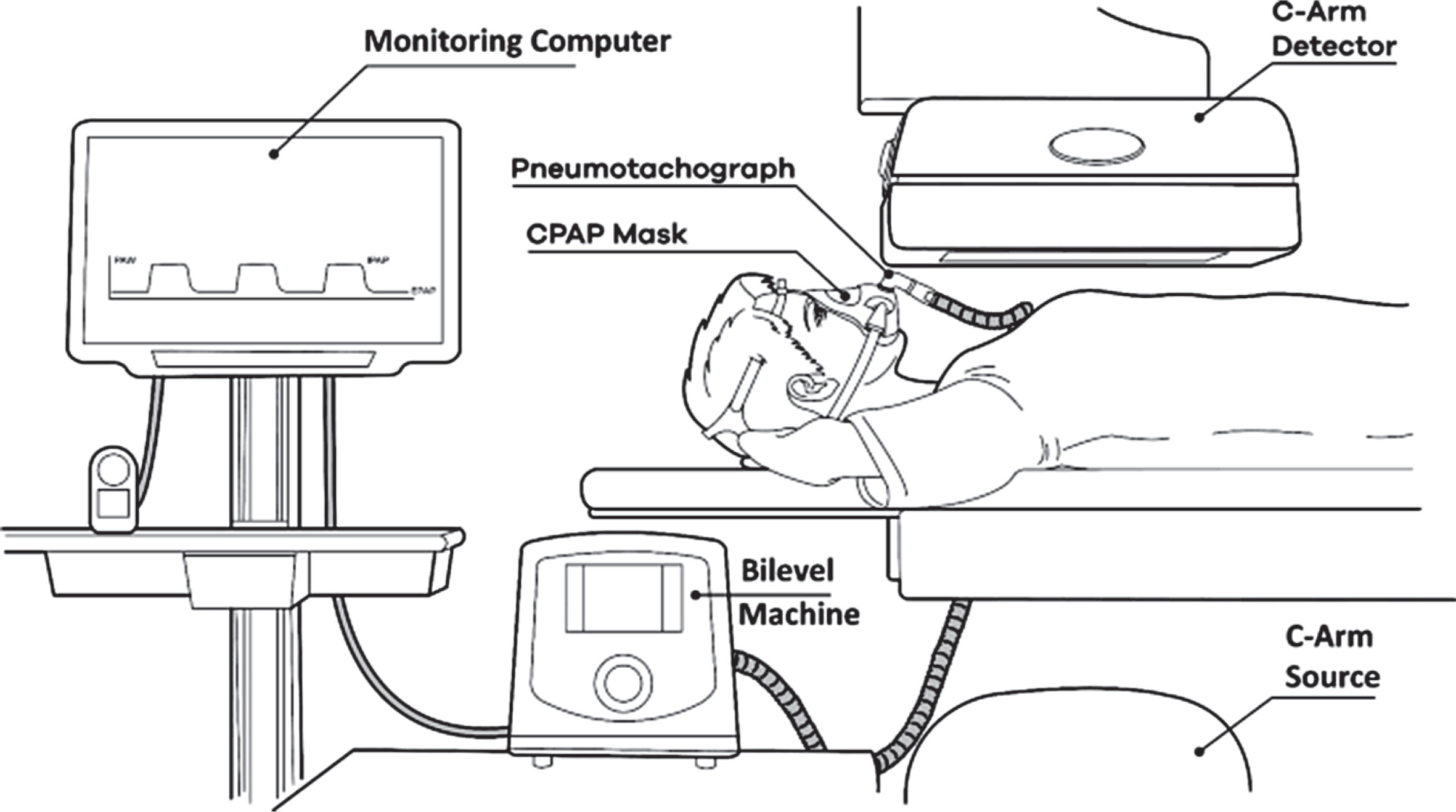
The abstracts were evaluated and assessed according to the following criteria: the papers were written in English, the studies comprised XV imaging outcomes and performances, clinical evaluations, studies that compared XV imaging to traditional imaging, clinical applications, image quality, and imaging aspects. Case investigations and case retrieval were both among the types of studies that were considered. Subsequently, the contents of the articles were evaluated to ascertain whether the imaging parameters of XV were confirmed. The studies and case collections that were not taken into consideration were those that had insufficient data and those with results that were inconclusive. The collections of entire publications were compiled based on the preliminary evaluation of titles and abstracts. Afterward, the results of the database search were used to eliminate duplicate research, and evaluations were carried out to determine whether the titles, abstracts, and overall conclusions of the publications met the selection conditions. The reviews of the reference collections of studies that were determined through the search process that was described previously were carried out, and further publications were found. A representation of the article preference approach can be found in Figure 1.
Electronic search
Searches were conducted on the electronic literature databases of ScienceDirect, Springer, PubMed/Medline, and Wiley Online Library to find papers relevant to all accessible dates. An original search strategy was created for PubMed and later enhanced for application in the other electronic databases. The primary search combined key XV imaging terms, such as lung imaging, with the following terms: lung evaluations, dynamic flow assessments, and vascular imaging.
Data extraction
From 2010 to 2024, electronic searches were conducted on MEDLINE (PubMed) and other databases. Mendeley was used for referencing. The entire paper was reviewed to ensure that it met the appropriate standards and that the data retrieval was appropriate. For each study, the following information was gathered: flow measurement parameters, imaging features, region distinction, differential investigation, air and blood flow evaluation, and any other relevant review process references. The most important findings, detection accuracy, sample size and features, year of publication, author name and country, and other pertinent data were retrieved from the included articles.
Data synthesis
The main objective was to research the imaging characteristics of XV imaging as well as the accuracy of dynamic flow measurement in air and blood analysis. Additionally, the capacity of this imaging technique to enable enhanced depiction of flow analysis was determined. As a part of the process of evaluating the performance and accuracy of the diagnosis, the findings of the imaging were evaluated and compared to the results obtained with traditional imaging techniques. Additional factors that were taken into consideration included changes in imaging methods and processing techniques.
Results
An electronic search utilising the above keywords produced 200 articles. Following whole text extraction and eliminating papers that did not fit the inclusion criteria, the findings were refined to 14 publications spanning 2010-2024. Studies were categorised according to technical factors, diagnostic efficacy, image quality, and flow analyses. The review consisted of 14 papers that were deemed eligible for inclusion. Four research studies demonstrated the capabilities of XV with spirometry [15,22-24]. Ten studies showed the performance analysis with various imaging techniques including CT, synchrotron imaging, particle image velocimetry, pitot flow meter, and phase contrast X-ray imaging [16,18-20,25-30]. Out of the investigations that were carried out, 5 centred on animals, with mice and rats being the primary subjects of investigation for the various research criteria [18,25-27,29]. Five other studies, on the other hand, engaged human patients and analysed data from clinical trials or medical observations to gain a better understanding of how the findings may apply to human health or disease [15,22-24,31]. In the other 4 investigations, XV analysis was utilised through the employment of simulations and animal models to refine and illustrate the application of the technique [19,20,28,30].
Most of the investigations that were conducted yielded findings that were substantially consistent with each other, with similar conclusions being taken from each study. In lung imaging, almost all the studies demonstrated that XV has been more efficient in recent years. These studies have shown that it can capture precise airflow dynamics and offer reliable respiratory assessments [15,22-29,31]. All the remaining research, on the other hand, has concentrated on the potential of the approach in vascular investigations, notably its use in determining and assessing blood flow. Studies have demonstrated that XV can provide useful insights into circulation patterns, which can assist in evaluating the health of the cardiovascular system and identifying potential problems with blood flow [18-20,30]. Broadly, the distinctive approach demonstrates potential in both pulmonary and vascular applications, which will enhance diagnostic skills in these extremely important segments of the body (Table 1).
Table 1
Comparative assessment of studies
| Author, year, country | Subjects | Techniques | Regions | Findings | Limitations |
|---|---|---|---|---|---|
| Dusting et al. 2020, USA [22] | Humans | Spirometry, computed tomography | Lungs | Extensive lung function data Early detection and assessment of diseases | Inadequate assessment of specific lung areas |
| Thurgood et al. 2016, Australia [25] | Animal | Synchrotron-based imaging | Lungs | Enhanced lung function analysis | Minimal analysis of apices region |
| Raoof et al. 2023, USA [23] | Humans | Spirometry | Lungs | Enhanced lung function analysis, stable ventilation phenotyping | Inadequate assessment of specific lung areas |
| Murrie et al. 2015, Australia [26] | Animal | Micro-tomography | Lungs | Extensive regional lung movement tracking | Soft tissue analysis |
| Werdiger et al. 2020, Australia [27] | Animal | Synchrotron-based imaging | Lungs | Enhanced lung function analysis, improved airflow dysfunction analysis | Reduced spatial resolution |
| Jamison et al. 2012, Australia [18] | Animal | Synchrotron-based imaging | Blood vessels | Accurate measurement of haemodynamics ex vivo | Low signal-to-noise ratio |
| Jamison et al. 2011, Australia [19] | Model | Particle image velocimetry | Blood vessels | Enhanced pulsatile blood flow imaging, accurate wall shear stress measurements | A limited number of frames, temporal resolution |
| Kirkness et al. 2023, USA [15] | Humans | Spirometry | Lung | Enhanced lung function analysis | Differential diagnosis of varying lung conditions and anomalies |
| Siddharthan et al. 2023, Australia [31] | Humans | Pitot tube flowmeter | Lung | Enhanced lung function analysis | Differential diagnosis of varying lung conditions |
| Murrie et al. 2016, Australia [28] | Model | Phase contrast X-ray imaging | Lung | Extensive lung function analysis, early detection and assessment of diseases | A limited number of frames, differential diagnosis |
| Bruorton et al. 2024, Australia [24] | Humans | Spirometry | Lung | Extensive lung function analysis | Differential diagnosis of lung regions |
| Goonan et al. 2018, Australia [29] | Animal | Particle image velocimetry | Lung | In vivo dynamic lung imaging, early detection and assessment of diseases | Inadequate assessment of lung |
| Dubsky et al. 2010, Australia [30] | Model | Computed tomography, particle image velocimetry | Blood vessel | Assessment of 3D blood flow-fields inside opaque vessels | Imaging setup, differential diagnosis |
| Dubsky et al. 2012, Australia [20] | Model | Computed tomography, particle image velocimetry | Blood vessel | Enhanced in vivo blood vessel measurement | Constant or periodic flow vessels |
Discussion
Spirometry
Spirometry is a prevalent and crucial pulmonary function test that assesses lung function by measuring the amount of air a person can inhale and exhale and the rate at which air is exhaled [32]. Spirometry provides useful information, including crucial measures such as forced expiratory volume in one second (FEV1) and forced vital capacity (FVC), which assesses the existence and severity of airflow obstruction or constraints [33]. Spirometry is an essential diagnostic tool for evaluating many obstructive lung diseases, such as asthma and COPD [34]. Spirometry enables practitioners to determine the degree of blockage, monitor disease progression, and evaluate the success of treatment [35]. Regular spirometry testing is essential for enhancing the quality of life for patients with chronic respiratory illnesses. It helps guide therapeutic decisions, alter medications, and improve long-term management methods [36,37].
However, spirometry has difficulties in detecting dynamic airflow and lung region-specific disorders. While it monitors total air capacity and flow, it does not provide precise information regarding airflow distribution in individual lung regions [12,38]. Furthermore, spirometry is a static test that cannot detect real-time changes in airflow during regular breathing or exercise. This makes it less useful for assessing dynamic changes in lung function, such as those observed in asthma or during rapid movements [39]. As a result, spirometry may not fully capture the complexity of airflow patterns or reveal localised lung abnormalities, especially if the blockage is minor or intermittent. While spirometry offers useful information about overall lung function, it has limits in detecting subtle or region-specific abnormalities. As a result, more advanced imaging techniques may be required for a more thorough lung health assessment and to assist proper diagnosis and assistance.
Computed tomography imaging
In the field of lung imaging and vascular diseases, CT is an indispensable tool because of its capacity to produce cross-sectional images that are both high resolution and detailed [4,40]. It is possible to identify and evaluate diseases such as lung cancer, COPD, pulmonary embolism, and interstitial lung diseases with the use of CT in lung imaging [41-43]. To see blood vessels, CT angiography is the gold standard in the field of vascular imaging [44]. This technique enables the detection of vascular abnormalities, blockages, and aortic aneurysms. Using contrast agents, CT can provide a thorough depiction of both veins and arteries, which is helpful in the diagnosis and treatment [45,46]. Nevertheless, despite its many benefits, CT has some drawbacks in lung and vascular imaging, notably concerning the evaluation of flowing blood and air. Since CT generally produces static anatomical images, it is not possible for it to directly monitor real-time airflow in the lungs or evaluate lung function, such as gas exchange or airflow restriction [47,48]. Although CT is an effective tool for viewing channel anatomy in vascular imaging, it cannot measure dynamic blood flow characteristics such as velocity or pressure gradients if used [49,50]. CT is an effective diagnostic tool, but for the dynamic assessment of the flowing structures, further analysis is required to fully evaluate and depict the patient’s condition and diagnosis.
X-ray velocimetry – working principle
XV is a specialised imaging technique that uses fluoroscopic X-ray imaging to capture high-speed sequences of a moving object, such as lung tissue during breathing. It operates by evaluating pixel displacement between consecutive frames to quantify the velocity of tissue motion, allowing for quantitative measurement of lung ventilation [51,52]. This approach provides real-time tracking of small anatomical components within tissue, allowing for detailed motion analysis without the need for intrusive treatments [52].
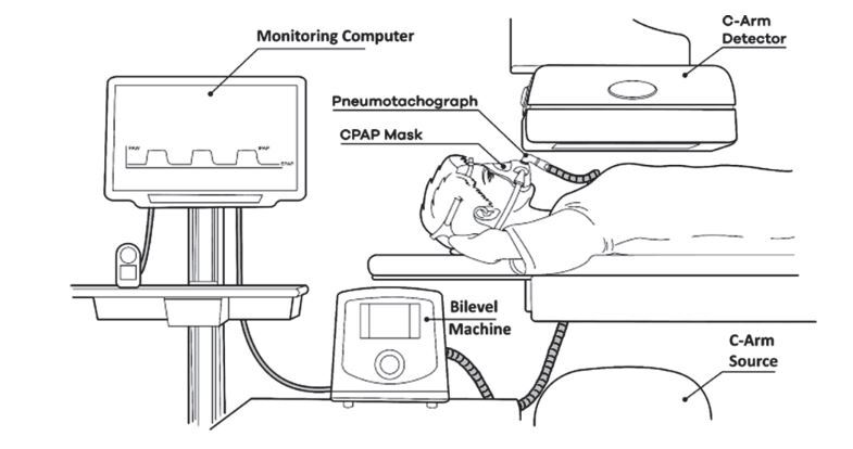
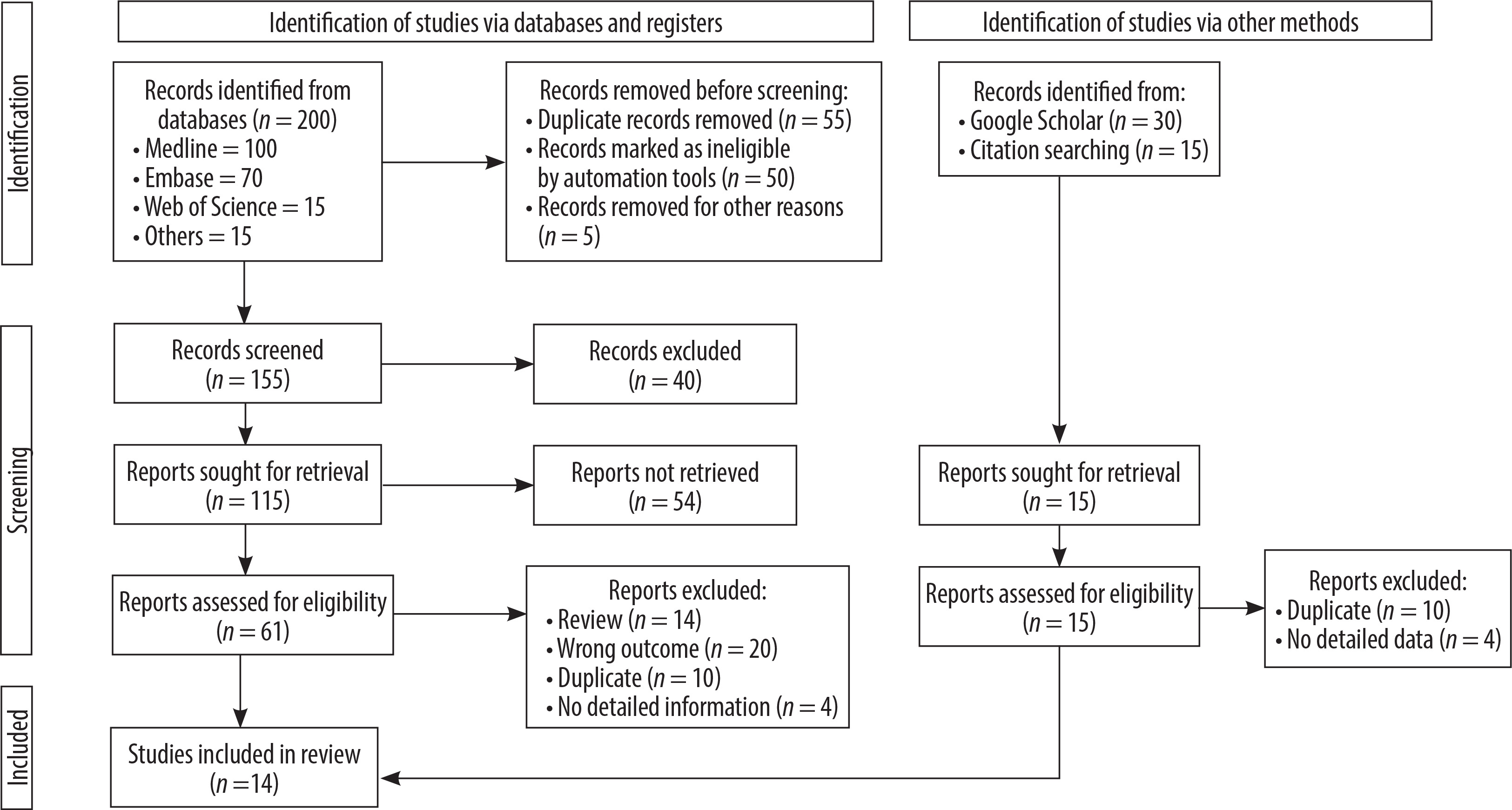
XV consists of 3 significant phases. Initially, a high-frame-rate fluoroscope gathers a continuous series of X-ray images of the target area. Second, image processing is accomplished over several frames using specialised software that detects and monitors characteristics, such as lung tissue markers or small blood vessels. Ultimately, the speed and direction of tissue movement are found utilising pixel displacement of these tracked properties between frames (Figure 2) [15,31,53].
It is an innovative functional imaging technique for the lungs. It tracks lung movements, calculates airway volumes, and assesses regional lung ventilation during natural breathing [31]. XV gives essential insight into lung mechanics and functioning by collecting detailed, real-time data on several respiratory parameters. This innovative diagnostic approach helps with accurate assessment of lung volumes, airflow patterns, and gas exchange efficiency [54].
Dusting et al. [22] demonstrated the peculiarities of XV about CT and spirometry by assessing radiotherapy patients’ lung functioning and working. XV analysis revealed local differences in associated breathing, providing rich lung function details that could not be observed using other modalities. XV showed immense capabilities when combined with novel techniques that could depict the measurement of lung function with varying imaging characteristics throughout different regions. It has also shown the potential to become a great diagnostic tool in the field of lung imaging [25]. Murrie et al. [26] highlighted that accurate insights into regional lung behaviour are made possible with the inclusion of XV, which improves the dynamic tracking of lung motion during breathing. By allowing for more precise evaluations of lung function, this development shows tremendous promise for the investigation of respiratory illnesses. Furthermore, by combining XV with micro-CT, new genetic and pharmacological treatments might be developed more quickly, which could advance personalised medicine and change the way a variety of respiratory conditions are treated. Recently, a study done on COPD patients by Raoof et al. [23] showed that spirometry tests with ventilation phenotyping employing XV are stable and repeatable. Both investigation and regular clinical practice can benefit from this method’s dependable, non-invasive radiography assessment of lung function. The use of XV in COPD management has the potential to improve patient monitoring and the development of tailored treatment plans, leading to better results and more progress in research.
XV analysis in addition to cystic fibrosis lung disease, might be useful in treating asthma, COPD, emphysema, and possibly lung cancer. When applied to the evaluation of lung function in a variety of disorders, this approach has the potential to yield priceless insights into the development of disease and the efficacy of treatment. Furthermore, XV can be an important tool for improving treatment strategies through the creation and evaluation of novel respiratory medicines [27]. A comparative study done by Kirkness et al. [15] implied that XV is a safe and highly effective method for many lung illnesses, making it excellent for use in clinical and research settings when considered with spirometry. Its great sensitivity makes it a good endpoint for clinical trials; it measures alterations in lung condition and enables precise evaluation of the magnitude of therapy effects. For this reason, XV is a vital resource for tracking the development of diseases and testing the efficacy of novel respiratory therapies. When compared to static radiography evaluations and traditional pulmonary function testing, XV precision in determining regional ventilation distribution yields superior insights. With XV, we can find out more about respiratory wellness because it gives us extensive, real-time information on pulmonary function in multiple areas. These improved capabilities make XV a potent tool for disease monitoring and tailored treatment plans [31]. Through the combination of phase contrast X-ray imaging and XV, it is possible to reliably monitor the movement of the lungs, hence enhancing the detection of disease regions that are regionalised during natural breathing. With this innovative combination, it is possible to conduct a precise evaluation of lung function in real time, which provides a better understanding of the dynamics of the respiratory system. It is possible to increase diagnosis accuracy and gain deeper insights into the progression of the disease by having the ability to track lung movements with a high level of detail, as illustrated in a study conducted on animal lungs by Murrie et al. [28].
A study conducted by Bruorton et al. [24] showed the immense role of XV in imaging children with normal lungs and children with cystic fibrosis. It provides in-depth and dynamic insights into the movements of the lungs, which enables accurate measurement of regional ventilation even in young children. This is especially helpful for detecting early lung abnormalities in cystic fibrosis, which can be difficult to detect with traditional methods because they may overlook small changes. Goonan et al. [29] highlighted the abilities of multisource XV in detection and analysis with improved spatial resolution and imaging features compared to single-source XV. It is also predicted that when it comes to advancing the development of an in vivo dynamic imaging system for human lungs, XV will play a significant part in shaping the future. Within the field of medical diagnostics, this system has the potential to become an indispensable diagnostic instrument for the early diagnosis of a variety of lung diseases, thereby satisfying a large demand.
Additionally, XV has proven its capability in assessing and monitoring blood flow, which provides vital insights into cardiovascular health and enhances diagnostic accuracy for a variety of vascular disorders. It has been established through research that it can measure intricate haemodynamics within the vasculature of real mammalian mammals. Using this technique, it is possible to see the dynamics of blood flow while also allowing for the application of pharmacological stimulation to change the conditions of the vessels. The precise measurement of blood flow in living organisms is still the ultimate objective of XV, although tremendous progress has been made in this region [18]. Jamison et al. [19] also showcased that XV is capable of imaging pulsatile blood flow in an in vitro model at physiologically realistic flow rates. Achieving in vivo velocimetry would make it possible to take velocity measurements with a high degree of accuracy, which would result in reliable information regarding wall shear stress. An increasing understanding of how haemodynamic factors influence vascular illnesses is made possible by this breakthrough, which is noteworthy.
Through the utilisation of XV, it is possible to measure 3-dimensional blood flow fields within opaque capillaries, which presents a distinct benefit for the investigation of cardiovascular disorders. When combined with tomographic imaging, XV makes it possible to conduct high-resolution, real-time visualisations of the dynamics of blood flow in living organisms. comprehension of haemodynamic parameters like blood velocity and wall shear stress, which are essential for evaluating cardiovascular diseases, is improved because of this combination. The diagnosis, risk assessments, and treatment methods for vascular illnesses could all be considerably improved with the implementation of this innovative imaging technique [20,30].
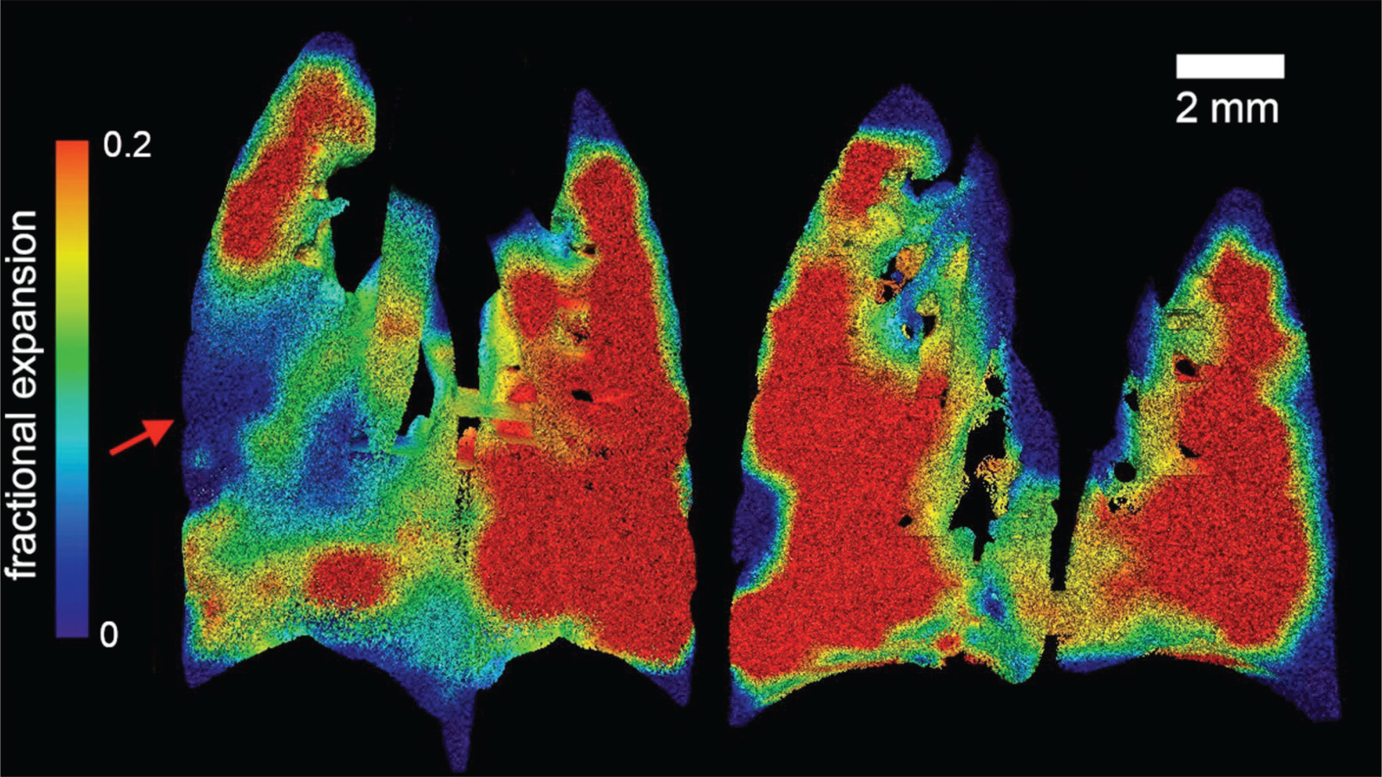
XV provides high-resolution, real-time, 3-dimensional blood flow analysis, and enhanced lung imaging and analysis, which enhances the knowledge of haemodynamics and respiratory functioning. However, it does have a few limitations. It has shown inadequate analysis of specific areas of the lung, mainly apical regions [22,23,25,29]. In the diagnosis of varying respiratory conditions with variations in interpretations [15,24,30] and the need for advanced synchrotron imaging systems and adequate image analysis mechanisms [19,28] it has also depicted its inability to differentiate constant or periodic flow vessels accurately [20].
Despite these limitations, High-resolution, real-time flow analysis is made possible by XV, which enables substantial breakthroughs in pulmonary and vascular imaging. By enhancing our understanding of respiratory dynamics and vascular difficulties, this technique has the potential to provide essential insights into flow patterns and haemodynamics. During the review process, a few challenges were encountered, including the limited number of published studies on XV imaging, inconsistencies in techniques and instrumentation, and variability in sample sizes and subject characteristics, which impacted the analysis and assessment processes.
Future developments in XV are anticipated to improve image resolution and accuracy, which will make it possible to conduct a more in-depth and accurate investigation of the dynamics of the lungs and the blood vessels. As a result of these enhancements, early identification, improved monitoring, and more customised medical care for pulmonary and cardiovascular conditions will be made possible. XV can potentially become a vital tool for enhancing diagnostic capacities and patient outcomes in those critical domains, provided that technological advancements take place (Figure 3).
Conclusions
XV appears to hold significant promise in the field of lung imaging as well as the assessment of blood flow, according to the majority of our investigations. This innovative imaging approach offers a high-resolution, real-time view of blood flow dynamics and respiratory function, providing essential insights into the health of the pulmonary and vascular systems. It is possible to gain a more precise understanding of haemodynamics, which includes aspects like blood velocity and wall shear stress, because of the capability of XV to measure blood flow in 3 dimensions. The ability to capture comprehensive patterns of airflow and lung function is a significant benefit in lung imaging because it assists in diagnosing and monitoring respiratory illnesses. Furthermore, the utilisation of XV in the field of vascular imaging boosts the capability to investigate irregularities in blood flow, which is an essential component in the diagnosis and treatment of cardiovascular disorders. The uses of XV in both domains may change early diagnosis, treatment planning, and disease monitoring as research continues to advance. This will ultimately lead to improvements in patient care and results.