

Introduction
This article is a narrative review centred on contrast-enhanced ultrasound (CEUS) in liver oncology, with emphasis on (i) focal liver lesion characterisation in chronic liver disease, (ii) assessment of residual viable tumour after locoregional therapies, and (iii) interventional guidance and intraprocedural decision support. The literature was selected to prioritise peer-reviewed clinical studies, consensus/technical reviews, and major translational reports published predominantly within the last 5 years, supplemented by earlier foundational work where it remains directly relevant.
Quantitative analysis in CEUS
A primary development in CEUS is the transition from qualitative pattern recognition [1-3] toward the use of quantitative, reportable parameters. Semi-quantitative or quantitative descriptors of enhancement – including wash-in slope, time to peak, peak intensity, and area under the curve – are provided through time – intensity curve (TIC) analysis and dynamic CEUS (DCE-CEUS). In liver oncology, these parameters are considered advantageous because the vascular phenotype may be reflected and longitudinal comparisons after therapy can be supported, provided that acquisition and post-processing are sufficiently standardised [ 4,5].
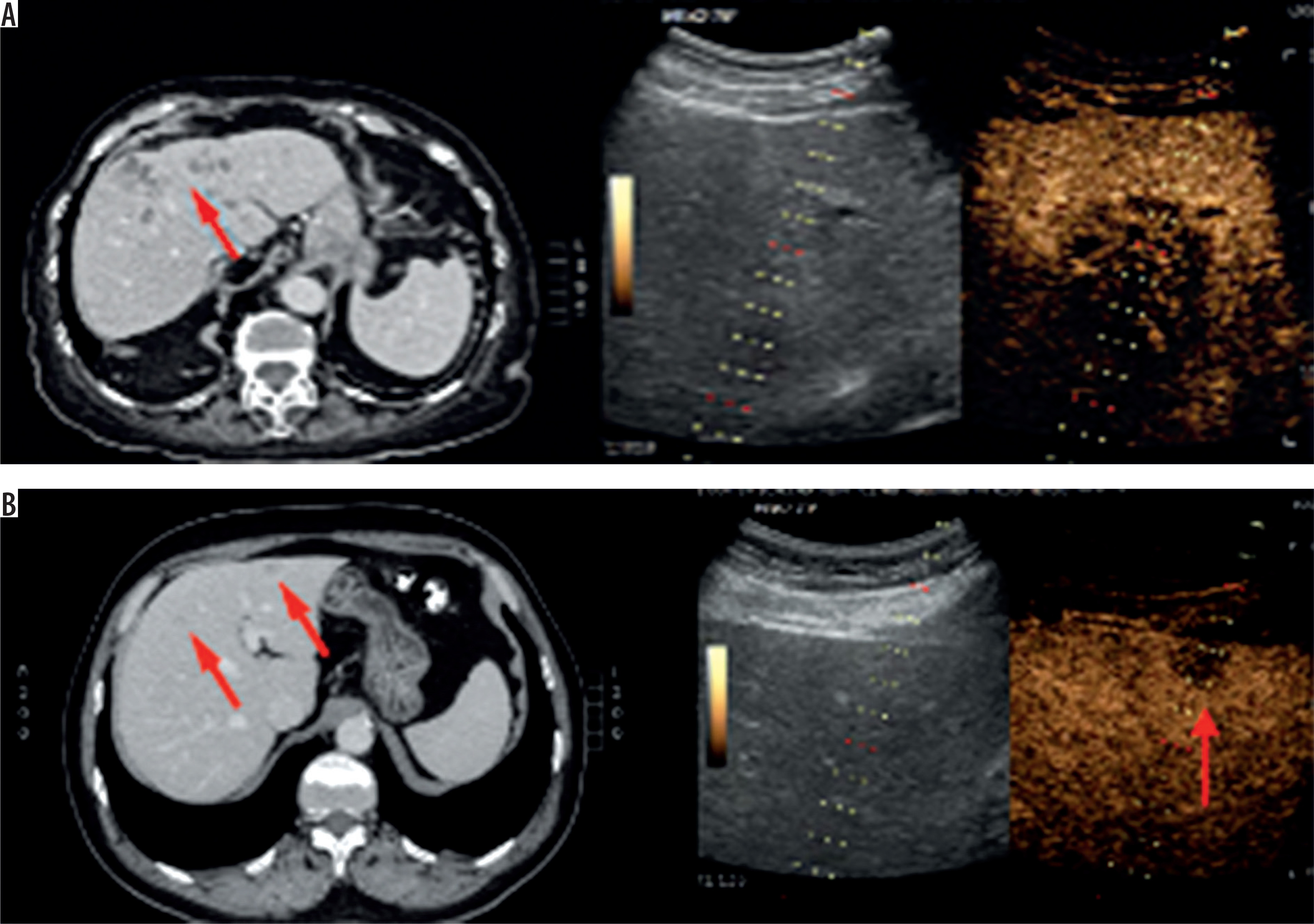
In the setting of locoregional therapy, such as ablation or embolisation, imaging evaluates whether residual viable tumour warranting early retreatment persists or if complete devascularisation has been achieved. The conspicuity of enhancing viable components within treated areas is improved by CEUS (Figure 1), which can be performed repeatedly at the point of care. Comparative diagnostic performance data regarding focal liver lesion characterisation are summarised in Table 1 [3].
Table 1
Data synthesis: The comparative analysis across both liver and renal studies demonstrates the superior diagnostic profile of CEUS. In liver imaging, CEUS significantly outperformed CT/MRI in terms of sensitivity and accuracy (p < 0.0001). Similarly, in the evaluation of unclear renal masses, CEUS maintained an exceptional sensitivity (99.1%) and a higher specificity (80.5%) compared to CT (47.4%). The high negative predictive value (NPV) observed in renal assessments (94.3%) indicates CEUS as a key tool for the non-invasive exclusion of malignancy, especially where cross-sectional imaging results remain indeterminate
Within the scope of treatment monitoring, two approaches have been proposed: size-based and viability-based response assessments. While RECIST 1.1 remains a widely utilised standard across solid tumours, size reduction is often observed later than the biologic response following locoregional therapies in liver oncology [6]. Therefore, enhancement-based concepts focusing on viable tumour are better aligned with the clinical requirement for early detection of residual activity. In this context, CEUS is viewed as conceptually consistent with enhancement-focused response frameworks (e.g. mRECIST/EASL) because persisting arterial hyperenhancement within treated lesions or ablation zones can be depicted, thereby supporting timely retreatment decisions [4,5].
Despite these advantages, quantitative CEUS remains sensitive to acquisition settings, vendor-specific processing, motion artifacts, ROI placement, and heterogeneous background liver parenchyma, particularly in cases of cirrhosis. Unless harmonised protocols and transparent reporting of technical parameters are established, quantitative results remain difficult to generalise across different centres and devices, which limits their application as trial-grade endpoints.
3D-CEUS and volumetric assessment
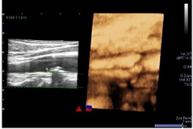
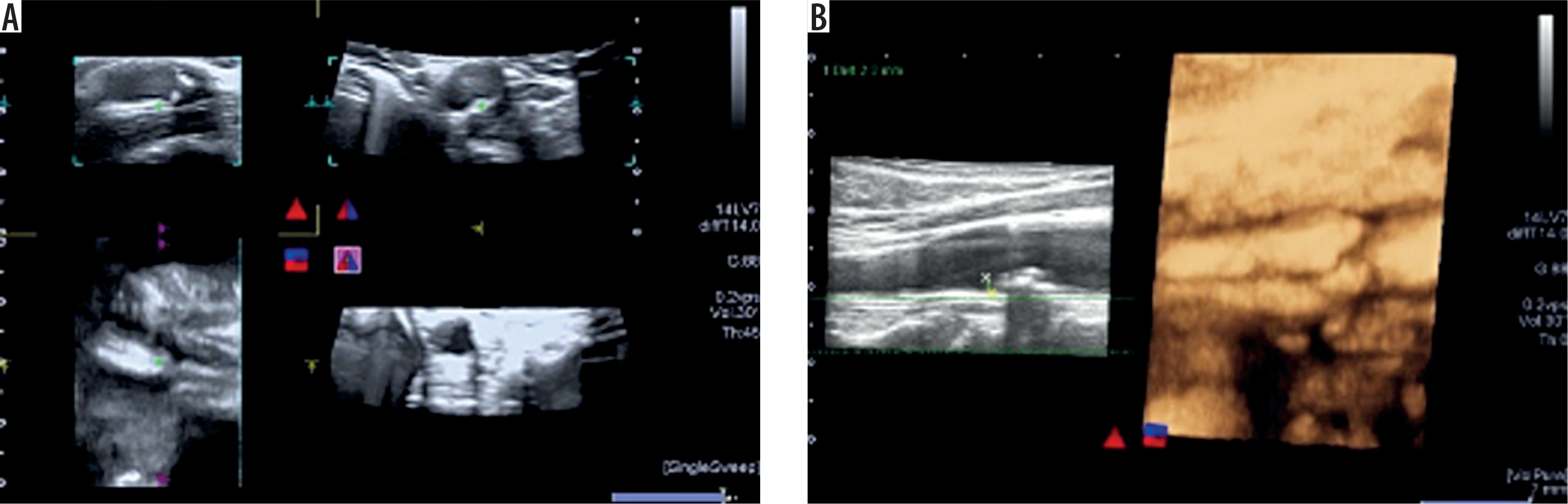
Heterogeneity or extent may be underestimated by two-dimensional CEUS when lesions are irregular, multifocal, or partially visualised, particularly within post-treatment beds. Sampling bias is addressed by 3D-CEUS and volumetric approaches, which enable the evaluation of enhancing volume instead of single-plane enhancement (Figure 2) [1].
Figure 2
Volumetric image – contrast-enhanced ultrasound. Carotid arteries. A) Orthogonal imaging. B) 4D rendering
In the context of post-ablation monitoring, this approach is particularly relevant for ablative margin (AM) assessment and the early prediction of local tumour progression. The evidence comparing 2D and 3D CEUS-derived approaches after RFA is summarised in Table 2 [7].
Table 2
2D vs. 3D assessment in post-ablation monitoring 3D-contrast-enhanced ultrasound (CEUS)/fusion imaging significantly outperformed 2D-CEUS by reducing registration time (5.0 vs. 9.3 min) and eliminating the sampling errors of single-plane scans. While 2D-CEUS misclassified 26% of insufficient margins as “safe,” 3D volumetric assessment achieved 100% sensitivity in detecting residual tumors, whereas 2D imaging reported only 55.6% (p = 0.019)
Routine adoption remains limited by practical barriers, including availability, acquisition time, breath-hold constraints, motion artifacts, and variability in post-processing. The clinical impact of 3D-CEUS is expected to depend upon integration with automated segmentation, longitudinal registration, and standardised reporting, rather than on volumetrics alone [1,7-9].
Fusion imaging and procedure guidance
Real-time ultrasound (including CEUS) is integrated with pre-procedural computed tomography (CT) or magnetic resonance imaging (MRI) through fusion imaging [10]. In the field of liver oncology, lesion localisation is improved, the targeting of lesions with poor B-mode visibility is enabled, and more confident biopsy or ablation is supported [11,12]. Functional information is added by CEUS through the highlighting of viable vascularity and the delineation of residual enhancement within treated zones.
Mapping is conducted using cross-sectional imaging; subsequently, targeting is facilitated by fusion-assisted navigation, and immediate assessment of residual viability is provided via CEUS to determine if additional treatment is required. A synthesis of clinical utility and technical performance metrics for fusion workflows is summarised in Table 3 [10].
Table 3
Clinical synthesis: contrast-enhanced ultrasound (CEUS)-computed tomography (CT)/magnetic resonance imaging (MRI) fusion proved to be a promising alternative in a diagnostic process. While standard CT/MRI was insufficient in 12% of cases, adding CEUS fusion identified additional lesions and caused immediate therapeutic strategy modifications. Furthermore, real-time microvascular imaging enabled a definitive diagnosis in 15% of patients with atypical CT/MRI enhancement patterns. Despite the higher registration success of ultrasound (US)-US fusion (93.8%), its low applicability rate (61.1%) due to poor B-mode visibility makes multimodal fusion a superior solution, offering 99.7% applicability and 99.3% technical efficacy
However, fusion benefits are found to be heterogeneous and case-dependent. Outcomes can be affected by registration errors, liver deformation due to respiration and probe pressure, and operator dependence, all of which should be explicitly acknowledged. Endpoints reflecting clinical utility – including procedural success, duration, complication rates, and the frequency with which intraprocedural CEUS evokes a change in therapy (e.g. additional passes, alternative approaches, or escalation to another modality) – should be prioritised in future comparative studies [10-12].
Artificial intelligence in CEUS: segmentation, radiomics, and prediction
Artificial intelligence (AI) has been integrated as a central component of contemporary imaging pipelines. In the context of CEUS, inter-observer variability in ROI selection – identified as a major source of noise in TIC-based quantification – may be reduced through automated segmentation [13].
Beyond segmentation, high-dimensional features can be extracted from CEUS videos and parametric maps via radiomics [14] and machine learning [15] to support lesion classification, risk stratification, and the prediction of recurrence or treatment response [16,17]. Representative deep learning studies and validation setups are summarised in Table 4 [13,17,18].
Table 4
Summary of deep learning models for liver lesion assessment. Because the objectives of these studies vary – ranging from lesion classification to recurrence prediction – different metrics were used to evaluate the performance of each method. The distinction between internal and external validation is also emphasized to show how the models perform both in their original clinical settings and across different hospitals. For segmentation tasks, the Intersection over Union (IoU) was used to measure the overlap between the artificial intelligence (AI)-generated outlines and the manual masks created by an expert. Finally, the “Reference” column indicates the established gold standard used to verify the accuracy of each AI model
Concurrently, AI is observed to be particularly vulnerable to domain shift in CEUS, resulting from heterogeneity in vendor processing, contrast dosing, presets, and acquisition protocols. Many published models remain restricted to single-centre cohorts and retrospective validation, whereby apparent performance is often inflated and transportability is limited. Clinical adoption will necessitate harmonised acquisition, robust feature stability assessment, external validation across multiple devices and centres, and prospective evaluation demonstrating management-changing benefits rather than retrospective discrimination alone [17-19].
Molecular CEUS and targeted microbubbles
Molecular CEUS goes beyond standard perfusion imaging by using microbubbles that bind to specific biomarkers, such as those involved in angiogenesis [19,20]. In liver oncology, this allows us to identify a tumour’s biological features and monitor its response at a molecular level [21,22].
Although the technology is promising, it has not reached routine clinical use yet. This is mainly due to strict regulatory hurdles, a lack of standardised agents, and the ongoing need to prove that it offers a real advantage over conventional CEUS, CT, or MRI [19-22].
Moreover, recent studies show that using novel ultrasound contrast agents – nanospheres – can widely enhance CEUS capabilities. Their size (under 400 nm) is considerably smaller than conventional microbubbles (> 1 μm), thus allowing them to move outside the blood vessel and into the tumour interstitially [23,24].
However, nanospheres tend to present poor selectivity in vivo after intervenous administration. This problem is overcome by the use of tumour-specific antibodies conjugated with nanospheres, which is a complex and treacherous process [23].
This approach can be particularly significant for pancreatic ductal adenocarcinoma (PDAC). Unlike many other tumours, PDAC is typically hypovascular and surrounded by a dense fibrotic stroma, which often renders conventional microbubbles ineffective. Targeted nanospheres can move past these vascular limitations, flowing directly into the pancreatic tumour matrix [23]. Such a method could allow for staging of small, otherwise inconspicuous pancreatic lesions.
Another study explored the implementation of nanospheres for facilitating small cell lung cancer imaging, proving that on the mouse model such an approach can translate into significantly higher AUC and peak intensity values [24].
UTMD and theranostics
Ultrasound-targeted microbubble destruction (UTMD) utilises acoustic energy to disrupt microbubbles [25]. This process creates a temporary increase in permeability, which facilitates the enhanced local delivery of therapeutics. Within the field of oncology, imaging is used to confirm perfusion and target characteristics, while controlled insonation drives localised delivery or works in synergy with ablative strategies. Successful clinical translation, however, requires meticulous control over acoustic parameters, dosing, and safety monitoring to minimise off-target effects and ensure reproducibility. Beyond UTMD, additional therapeutic ultrasound strategies – such as combining focused ultrasound with immunotherapy – are currently under clinical investigation. A summary of ongoing trials for these applications is provided in Table 5.
Table 5
Overview of ongoing clinical trials investigating the therapeutic applications of ultrasound
UTMD evolves from the sonoporation concept, which originated from foundational studies on the mechanical index (MI) and its application in CEUS. The MI serves as a standardised estimate of the maximum peak negative acoustic pressure within the tissue, scaled by the square root of the centre frequency; fundamentally, it quantifies the likelihood of microbubble cavitation [26]. From a clinical standpoint, this creates a vital distinction in ultrasound application: while a low MI is essential for stable, non-destructive real-time visualisation, escalating the acoustic intensity facilitates microbubble rupture, i.e. the very mechanism that enables targeted therapeutic delivery.
Empirical research, particularly studies involving the Sonazoid contrast agent, demonstrates that microbubble dissolution begins at moderate pressures, typically within an MI range of 0.4 to 1.0 [27]. However, true inertial cavitation – characterised by violent bubble collapse – is only triggered when the MI exceeds 1.0. This threshold is critical because the resulting disintegration of microbubbles provides the mechanical basis for sonoporation, defined as the use of ultrasound in the presence of cavitation nuclei to induce plasma membrane perforation [28].
In this context, the injected microbubbles serve as the essential cavitation nuclei. Because biological tissues and fluids naturally lack stabilised gas bodies within, they are inherently resistant to cavitation at diagnostic energy levels. By introducing synthetic microbubbles into the circulation, we provide “seeds” or nuclei that concentrate acoustic energy. These gas-filled cores oscillate and eventually collapse under ultrasound exposure, lowering the energy threshold required to generate the mechanical forces necessary for membrane permeabilisation.
In practical terms, while an MI of approximately 0.4 may be viewed as a “cellular massage” that gently disturbs the membrane, an MI exceeding 1.0 should be considered a “cellular puncture”. This localised perforation is a primary goal because it transiently disrupts membrane integrity to allow the uptake of exogenous drugs or genetic material. However, the success of this approach relies on a narrow safety window. Live microscopy has shown that sonoporation sites are dynamic; small, site-specific pores can be successfully repaired by the cell’s innate defence mechanisms – such as calcium-dependent resealing – often within 60 seconds. Conversely, excessive acoustic energy may lead to pores larger than 100 μm2 or severe disruption of the actin cytoskeleton, resulting in irreversible cell lysis or apoptosis. Thus, precisely calibrating the MI is essential to maximise delivery efficiency while maintaining cellular viability.
The clinical viability of this mechanical approach is best exemplified by recent breakthroughs in treating glioblastoma (GBM). While the blood-brain barrier (BBB) remains a formidable obstacle for most chemotherapeutic agents, magnetic resonance-guided focused ultrasound (MRgFUS) has successfully leveraged sonoporation to transiently disrupt this barrier. Clinical data have demonstrated that this technique can achieve a 7.7-fold increase in temozolomide (TMZ) concentration within sonicated areas compared to untreated tissue, notably without inducing long-term neurological complications.
It is important to emphasise, however, that these landmark results were achieved under MRI guidance, which provides excellent anatomical precision but lacks the real-time temporal resolution to monitor microbubble cavitation dynamics directly. The current challenge lies in translating these therapeutic successes from the MRI suite to ultrasound-guided platforms, such as CEUS [29].
UTMD and sonobiopsy
While sonoporation is predominantly utilised for the “inward” delivery of therapeutics, its potential for the “outward” release of tumour-specific biomarkers into the bloodstream is increasingly recognised [30]. In the field of neuro-oncology, this transition allows for a move from purely therapeutic interventions toward a comprehensive diagnostic and monitoring paradigm. The safety profile of this approach has been demonstrated in clinical trials involving patients with Alzheimer’s disease, where it was shown that biological barriers can be opened across multiple brain regions without causing permanent injury [31].
This emerging diagnostic method, termed sonobiopsy, utilises focused ultrasound and microbubbles to facilitate “two-way trafficking” between brain tissue and the peripheral vasculature. By briefly opening the blood-brain barrier, biomarkers such as circulating tumour DNA and mRNA are released into the plasma for collection. It must be noted that the first human study on this technique, published in 2023, was conducted on a limited cohort of only five participants; however, the results confirmed the feasibility of enriching biomarkers in the bloodstream [32]. This technique addresses the primary limitations of conventional liquid biopsies, where brain-derived markers are often too diluted or restricted by the blood-brain barrier to be detected. By enhancing the sensitivity of blood tests in a non-invasive and repeatable manner, sonobiopsy allows for the tracking of tumour changes without the risks associated with invasive brain surgery. However, this technique remains insufficiently investigated and requires further research.
Limitations and standardisation roadmap
Standardisation is deemed critical for the successful translation of CEUS innovations into routine liver oncology practice. Key barriers, such as vendor-dependent quantification, inconsistent acquisition protocols, variable operator expertise, and limited multi-centre prospective evidence, are frequently identified as obstacles to widespread implementation.
For quantitative CEUS, the inclusion of minimum reporting items is required; these should encompass system configurations (including mechanical index), contrast agent specifications and dosage, timing and breath-hold strategies, ROI/segmentation methodologies, motion management, reference standards, and reproducibility metrics. Regarding AI-based applications, the transparent reporting of preprocessing, calibration, external validation, and robustness across multiple vendors is considered essential.
A pragmatic roadmap is proposed, incorporating harmonised protocols, shared benchmark datasets, and prospective studies anchored in management-changing endpoints, such as retreatment decisions based on the early assessment of residual viability [1,2,4,5,15,16,19].
Discussion
The clinical value of CEUS in liver oncology is increasingly defined not by the replication of CT or MRI, but by advantages that are found difficult to achieve with other modalities [1-3]. These include real-time assessment of vascularity, repeatability without ionising radiation, and feasibility at the point of care. These strengths are identified as most critical in focal liver lesion characterisation in the context of chronic liver disease, early detection of residual viable tumour following locoregional therapies, and interventional guidance when rapid, iterative imaging is required.
Current advances in quantification, volumetrics, and AI are interpreted as systematic efforts to convert these pragmatic advantages into standardised outputs stable enough to influence clinical decisions and trial endpoints [1,2,4].
Objective enhancement descriptors are offered by quantitative CEUS, but the translation into clinically actionable parameters remains incomplete. Reproducibility is frequently compromised by cirrhosis-related heterogeneity, motion artifacts, ROI dependence, and vendor-specific variability. Consequently, parameters that are not only measurable but decision-relevant are prioritised, particularly those enabling the early identification of residual viability that triggers retreatment [5,6]. In this context, enhancement-focused response concepts are considered more aligned with the clinical question than size-only assessments, while RECIST 1.1 is maintained as an important cross-tumour reference standard for general oncologic trials [6].
Sampling bias may be reduced and post-ablation margin evaluation improved through 3D-CEUS and volumetric assessment, although their impact is expected to depend on workflow integration and automation [1,7]. Similarly, procedural reliability is enhanced by fusion imaging, which provides immediate post-treatment verification; however, the benefit is noted to be case-dependent and limited by registration errors and operator dependence [10-12]. Meaningful evidence is therefore expected to emphasise procedural outcomes and management changes rather than surrogate technical metrics alone [7,10].
AI is recognised as a highly visible direction, but the risk of overestimating its clinical readiness is acknowledged. Domain shift is identified as a core problem in CEUS due to vendor and protocol variability, and a lack of robust external validation is observed in many current studies. Future work must prioritise harmonised acquisition, multi-centre validation, and the prospective demonstration that AI-assisted CEUS improves clinical decisions and patient outcomes [14-19].
Finally, a compelling theranostic narrative is offered by molecular CEUS and UTMD, although these technologies require further research. Their translation is seen to depend on rigorous safety characterisation, standardisation of targeted agents, regulatory progress, and trials demonstrating incremental benefit beyond established systemic and locoregional strategies [19-22].
Conclusions
CEUS is considered to have turned into a clinically valuable modality in liver oncology by enabling real-time, repeatable assessment of tumour vascularity and immediate post-procedural evaluation of residual viability. Over the next decade, progress is expected to be judged primarily by management-changing benefit – particularly the earlier and more reliable detection of residual viable tumour after locoregional therapy – rather than by incremental improvements in standalone diagnostic performance [1,2,4,5].
Near-term clinical impact is predicted to arise from pragmatic integration: standardised quantitative protocols, workflow-optimised fusion imaging for selected interventional cases, and AI tools that reduce operator dependence while remaining robust across vendors and centres. In contrast, molecular CEUS and UTMD are represented as promising longer-horizon directions that will require rigorous safety evaluation and clinical trials demonstrating incremental benefit beyond established liver oncology strategies. Overall, harmonisation, external validation, and the use of prospective, clinically actionable endpoints are considered essential to enable wider adoption and reproducible patient benefit [7,9-11,14-22].

