

TECHNOLOGY AND CONTRAST MEDIA / ORIGINAL PAPER
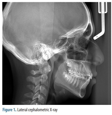
Artificial intelligence as a diagnostic tool using lateral cephalometric imaging
1
Mohammed Bin Rashid University of Medicine and Health Sciences, Dubai Healthcare City, Dubai, United Arab Emirates
Submission date: 2025-08-04
Final revision date: 2026-03-25
Acceptance date: 2026-03-26
Publication date: 2026-10-29
Corresponding author
Amani Idres
Mohammed Bin Rashid University of Medicine and Health Sciences, Dubai Healthcare City, Dubai, United Arab Emirates, 66566
Mohammed Bin Rashid University of Medicine and Health Sciences, Dubai Healthcare City, Dubai, United Arab Emirates, 66566
Pol J Radiol, 2026; 91(1): 526-537
KEYWORDS
TOPICS
ABSTRACT
Purpose:
To evaluate the accuracy and reliability of a newly developed artificial intelligence (AI)-driven system in identifying cephalometric landmarks, comparing its performance to manual tracing by orthodontists.
Material and methods:
A total of 506 lateral cephalometric radiographs of patients aged 14 to 40 years were collected from Dubai Dental Hospital, United Arab Emirates. After applying exclusion criteria, 473 radiographs were included in the final analysis. Manual tracings were performed using Dolphin Imaging software, identifying key landmarks including Sella, Nasion, A-point, B-point, Menton, Glabella, and others. The AI-based cephalometric system (AI Ceph) was trained on these radiographs to locate the same landmarks. Its performance was compared to manual tracings to assess accuracy and reliability.
Results:
The AI Ceph system demonstrated high concordance with manual tracing. The Menton point showed the highest agreement (96.6%), while Nasion had the lowest (86.6%). The majority of landmark deviations were within 2 mm, the predefined clinically acceptable range, while a small proportion exceeded this threshold and were classified as discordant (> 2 mm) according to the prespecified binary criterion. Sensitivity was above 80% for all landmarks, except Nasion (72.7%) and A-point (78.8%). A statistically significant improvement in concordance after model retraining was observed only for the Glabella landmark (p < 0.0213).
Conclusions:
The AI system provided accurate and reliable cephalometric landmark identification. While most deviations fell within the clinically acceptable 2-mm tolerance, landmarks exceeding this threshold were clearly identified and quantified using the binary discordance classification (> 2 mm), ensuring transparent reporting of all errors.
To evaluate the accuracy and reliability of a newly developed artificial intelligence (AI)-driven system in identifying cephalometric landmarks, comparing its performance to manual tracing by orthodontists.
Material and methods:
A total of 506 lateral cephalometric radiographs of patients aged 14 to 40 years were collected from Dubai Dental Hospital, United Arab Emirates. After applying exclusion criteria, 473 radiographs were included in the final analysis. Manual tracings were performed using Dolphin Imaging software, identifying key landmarks including Sella, Nasion, A-point, B-point, Menton, Glabella, and others. The AI-based cephalometric system (AI Ceph) was trained on these radiographs to locate the same landmarks. Its performance was compared to manual tracings to assess accuracy and reliability.
Results:
The AI Ceph system demonstrated high concordance with manual tracing. The Menton point showed the highest agreement (96.6%), while Nasion had the lowest (86.6%). The majority of landmark deviations were within 2 mm, the predefined clinically acceptable range, while a small proportion exceeded this threshold and were classified as discordant (> 2 mm) according to the prespecified binary criterion. Sensitivity was above 80% for all landmarks, except Nasion (72.7%) and A-point (78.8%). A statistically significant improvement in concordance after model retraining was observed only for the Glabella landmark (p < 0.0213).
Conclusions:
The AI system provided accurate and reliable cephalometric landmark identification. While most deviations fell within the clinically acceptable 2-mm tolerance, landmarks exceeding this threshold were clearly identified and quantified using the binary discordance classification (> 2 mm), ensuring transparent reporting of all errors.
REFERENCES (23)
1.
Allareddy V, Rengasamy Venugopalan S, Nalliah RP, Caplin JL, Lee MK, Allareddy V. Orthodontics in the era of big data analytics. Orthod Craniofac Res 2019; 22: 8-13.
2.
Ahmad Z, Rahim S, Zubair M, Abdul-Ghafar J. Artificial intelligence (AI) in medicine, current applications and future role with special emphasis on its potential and promise in pathology: present and future impact, obstacles including costs and acceptance among pathologists, practical and philosophical considerations. A comprehensive review. Diagn Pathol 2021; 16: 24. DOI: 10.1186/s13000-021-01085-4.
3.
Albarakati S, Kula K, Ghoneima A. The reliability and reproducibility of cephalometric measurements: a comparison of conventional and digital methods. Dentomaxillofac Radiol 2012; 41: 11-17.
4.
Alessandri-Bonetti A, Sangalli L, Salerno M, Gallenzi P. Reliability of artificial intelligence-assisted cephalometric analysis. A pilot study. BioMedInformatics 2023; 3: 44-53.
5.
Ali A, Chandna AK, Munjal A. Accuracy and reliability of soft tissue landmarks using three-dimensional imaging in comparison with two-dimensional cephalometrics: a systematic review. J Indian Orthod Soc 2020; 54: 289-296.
6.
Alqahtani J, Alhemaid G, Alqahtani H, Abughandar A, AlSaadi R, Algarni I, et al. Digital diagnostics and orthodontic practice. J Healthc Sci 2022; 2: 112-117.
7.
Alsubai S. A critical review on the 3D cephalometric analysis using machine learning. Computers 2022; 11: 154. DOI: 10.3390/computers11110154.
8.
Bahaa K, Noor G, Yousif Y. The artificial intelligence approach for diagnosis, treatment and modelling in orthodontic. In: Naretto S, editor. Principles in contemporary orthodontics. London: InTech; 2011, p. 451-492.
9.
Cunningham C, Scheuer L, Black S. Developmental juvenile osteology. 2nd ed. London: Academic Press; 2016.
10.
Mohan A, Sivakumar A, Nalabothu P. Evaluation of accuracy and reliability of OneCeph digital cephalometric analysis in comparison with manual cephalometric analysis – a cross-sectional study. BDJ Open 2021; 7: 22. DOI: 10.1038/s41405-021-00077-2.
11.
Jayachandran S. Digital imaging in dentistry: a review. Contemp Clin Dent 2017; 8: 193-194.
12.
Dilsizian SE, Siegel EL. Artificial intelligence in medicine and cardiac imaging: harnessing big data and advanced computing to provide personalized medical diagnosis and treatment. Curr Cardiol Rep 2014; 16: 441. DOI: 10.1007/s11886-013-0441-8.
13.
Ahmed N, Abbasi MS, Zuberi F, Qamar W, Halim MSB, Maqsood A, Alam MK. Artificial intelligence techniques: analysis, application, and outcome in dentistry – a systematic review. Biomed Res Int 2021; 2021: 9751564. DOI: 10.1155/2021/9751564.
14.
Gill SS, Xu M, Ottaviani C, Patros P, Bahsoon R, Shaghaghi A, et al. AI for next generation computing: emerging trends and future directions. Internet Things 2022; 19: 100514. DOI: 10.1016/j.iot.2022.100514.
15.
Liu J, Zhang C, Shan Z. Application of artificial intelligence in orthodontics: current state and future perspectives. Healthcare (Basel) 2023; 11: 2760. DOI: 10.3390/healthcare11202760.
16.
Kamoen A, Dermaut L, Verbeeck R. The clinical significance of error measurement in the interpretation of treatment results. Eur J Orthod 2001; 23: 569-578.
17.
Chen SK, Chen YJ, Yao CCJ, Chang HF. Enhanced speed and precision of measurement in a computer-assisted digital cephalometric analysis system. Angle Orthod 2004; 74: 501-507.
18.
Erkan M, Gurel HG, Nur M, Demirel B. Reliability of four different computerized cephalometric analysis programs. EurJ Orthod 2012; 34: 318-321.
19.
Lindner C, Wang CW, Huang CT, Li CH, Chang SW, Cootes TF. Fully automatic system for accurate localisation and analysis of cephalometric landmarks in lateral cephalograms. Sci Rep 2016; 6: 33581. DOI: 10.1038/srep33581.
20.
Uysal T, Baysal A, Yagci A. Evaluation of speed, repeatability, and reproducibility of digital radiography with manual versus computer-assisted cephalometric analyses. European J Orthod 2009; 31: 523-528.
21.
Hwang HW, Park JH, Moon JH, Yu Y, Kim H, Her SB, et al. Automated identification of cephalometric landmarks: Part 2 – Might it be better than human? Angle Orthod 2020; 90: 69-76.
22.
Meriç P, Naoumova J. Web-based fully automated cephalometric analysis: comparisons between app-aided, computerized, and manual tracings. Turk J Orthod 2020; 33: 142-149.
23.
Bulatova G, Kusnoto B, Grace V, Tsay TP, Avenetti DM, Sanchez FJC. Assessment of automatic cephalometric landmark identification using artificial intelligence. Orthod Craniofac Res 2021; 24: 37-42.
Share
RELATED ARTICLE
| ISSN: | 1899-0967 |

We process personal data collected when visiting the website. The function of obtaining information about users and their behavior is carried out by voluntarily entered information in forms and saving cookies in end devices. Data, including cookies, are used to provide services, improve the user experience and to analyze the traffic in accordance with the Privacy policy. Data are also collected and processed by Google Analytics tool (more).
You can change cookies settings in your browser. Restricted use of cookies in the browser configuration may affect some functionalities of the website.
You can change cookies settings in your browser. Restricted use of cookies in the browser configuration may affect some functionalities of the website.