

Introduction
Numerous minimally invasive and invasive diagnostic techniques are used for the diagnosis of mediastinal and lung lesions, including image-guided percutaneous transthoracic biopsy, transbronchial biopsy, thoracoscopy, mediastinoscopy, and thoracotomy [1,2]. Among these, percutaneous transthoracic biopsy techniques, comprising core needle biopsy (CNB) and fine-needle aspiration biopsy (FNA), are established as safe and effective procedures with acceptable complication rates [3-5]. Percutaneous biopsy procedures can be performed under either ultrasound (US) or computed tomography (CT) guidance, depending on the lesion location. US-guided transthoracic biopsy is particularly suitable for lesions involving the mediastinum, pleura, or peripheral lung parenchyma [6]. This approach offers distinct advantages, including real-time imaging and the absence of ionizing radiation. On the other hand, CT guidance is the preferred modality for accessing centrally located intraparenchymal lesions that are beyond the reach of bronchoscopy [7].
Transthoracic biopsy is a well-established diagnostic technique for the identification of both tumoral lesions and non-tumoral processes in adults with low complication rates, which are manageable, particularly when compared to thoracotomy (1-2% mortality for open biopsy) or video-assisted thoracoscopic surgery (1% mortality) [8]. Pneumothorax and pulmonary hemorrhage are the most commonly encountered complications of image-guided percutaneous transthoracic biopsy, while air embolism and tumor seeding along the needle path are rare [2]. Despite their widespread use in adults, image-guided transthoracic biopsy procedures are less frequently applied in pediatric populations. This study aims to evaluate the efficacy, safety, and diagnostic performance of image-guided transthoracic CNB in children, based on a retrospective analysis of 40 pediatric patients.
Material and methods
Patients
This retrospective single-center study included 40 pediatric patients who underwent image-guided transthoracic CNB between May 2011 and January 2025. All patients who met this criterion during the study period were included, and no exclusion criteria were applied, thereby minimizing the risk of selection bias. Data on demographic characteristics, imaging modalities used for guidance, lesion size, pathological diagnoses, and procedure-related complications were collected. Complications were classified based on the guidelines for percutaneous needle biopsy established by the Society of Interventional Radiology (SIR) [9].
In all patients, the indication for transthoracic biopsy was established following multidisciplinary evaluation involving pediatric radiology, pediatric oncology, pediatric surgery, and pediatric infectious diseases. Transthoracic biopsy was selected when a definitive tissue diagnosis was considered essential for guiding treatment decisions and could not be obtained through less invasive diagnostic approaches or from an alternative, lower-risk biopsy site (e.g., peripheral lymphadenopathy).
Patient preparation
Coagulation parameters and a complete blood count were evaluated. Patients using antiplatelet or anticoagulant medications withheld them prior to the procedure, taking into account the elimination half-life of the active substance and in consultation with the prescribing clinician to ensure patient safety. The patient’s parents or legal guardians were informed in detail about the procedure and its potential complications, and written informed consent was obtained. This retrospective study received approval from the institutional research ethics board (decision number: 2025/47).
Technique
All procedures were performed by an experienced interventional radiologist under intravenous sedation provided by the anesthesia team. US guidance was preferred for mediastinal lesions and pleural-based or peripherally located lung lesions, whereas CT guidance was used for biopsies of centrally located lung parenchymal lesions. The coaxial technique was used for CT-guided biopsy procedures, while either the single-needle or coaxial technique was employed for US-guided biopsies. Patients were positioned in the supine, prone, or lateral decubitus position based on the lesion’s location in both types of procedures.
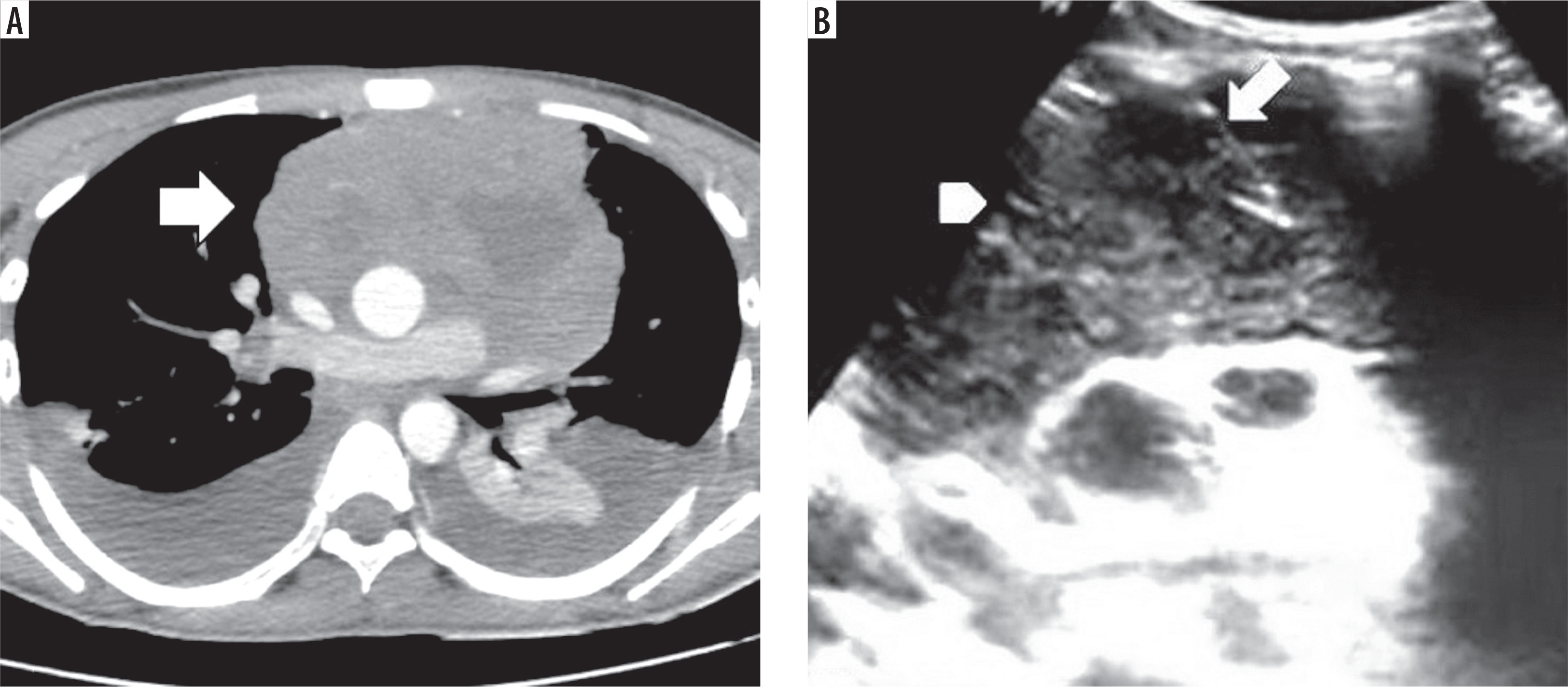
In US-guided procedures, real-time imaging guidance was performed using a C1-5-RS wideband convex array probe (Logiq P9, GE Healthcare, San Diego, Calif). The puncture site was chosen to ensure optimal access to the lesion while avoiding vascular structures. After disinfecting the area, local anesthesia was administered. Tissue samples were obtained using an 18- or 20-gauge semi-automatic Tru-Cut biopsy needle (Geotek Medical, Ankara, Turkey), with or without the use of a coaxial technique (Figure 1). Following the procedure, follow-up chest radiography was performed to evaluate for potential complications.
Figure 1
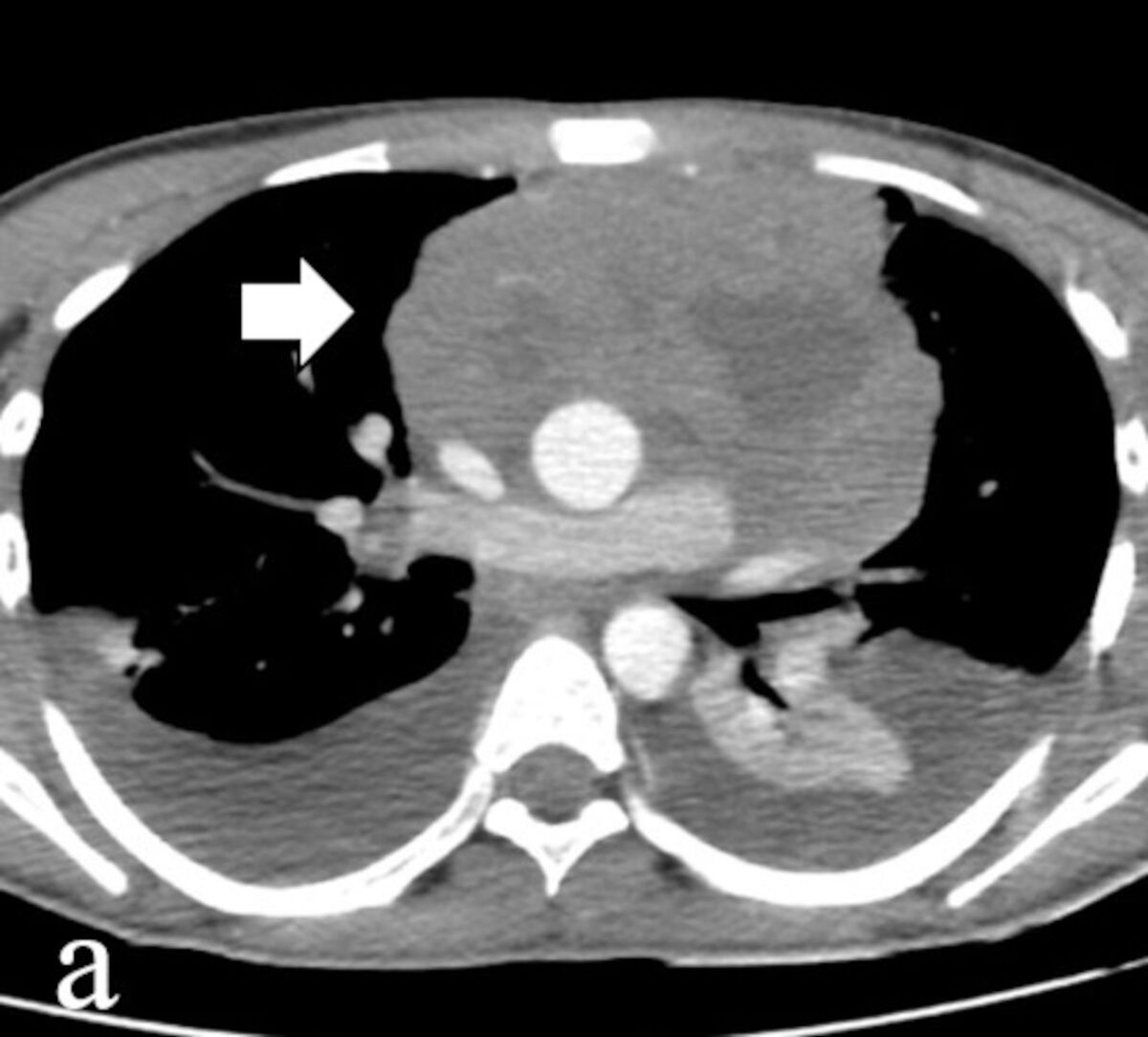
An axial contrast-enhanced CT scan of a 16-year-old male patient demonstrates a large mass in the anterior mediastinum (arrow) (A). The ultrasound (US) image obtained during US-guided percutaneous transthoracic biopsy shows a hypoechoic mass in the anterior mediastinum (arrowhead) with the biopsy needle clearly visible within the lesion (arrow) (B). No complications were observed during or after the procedure, and the pathological diagnosis was B-cell lymphoma
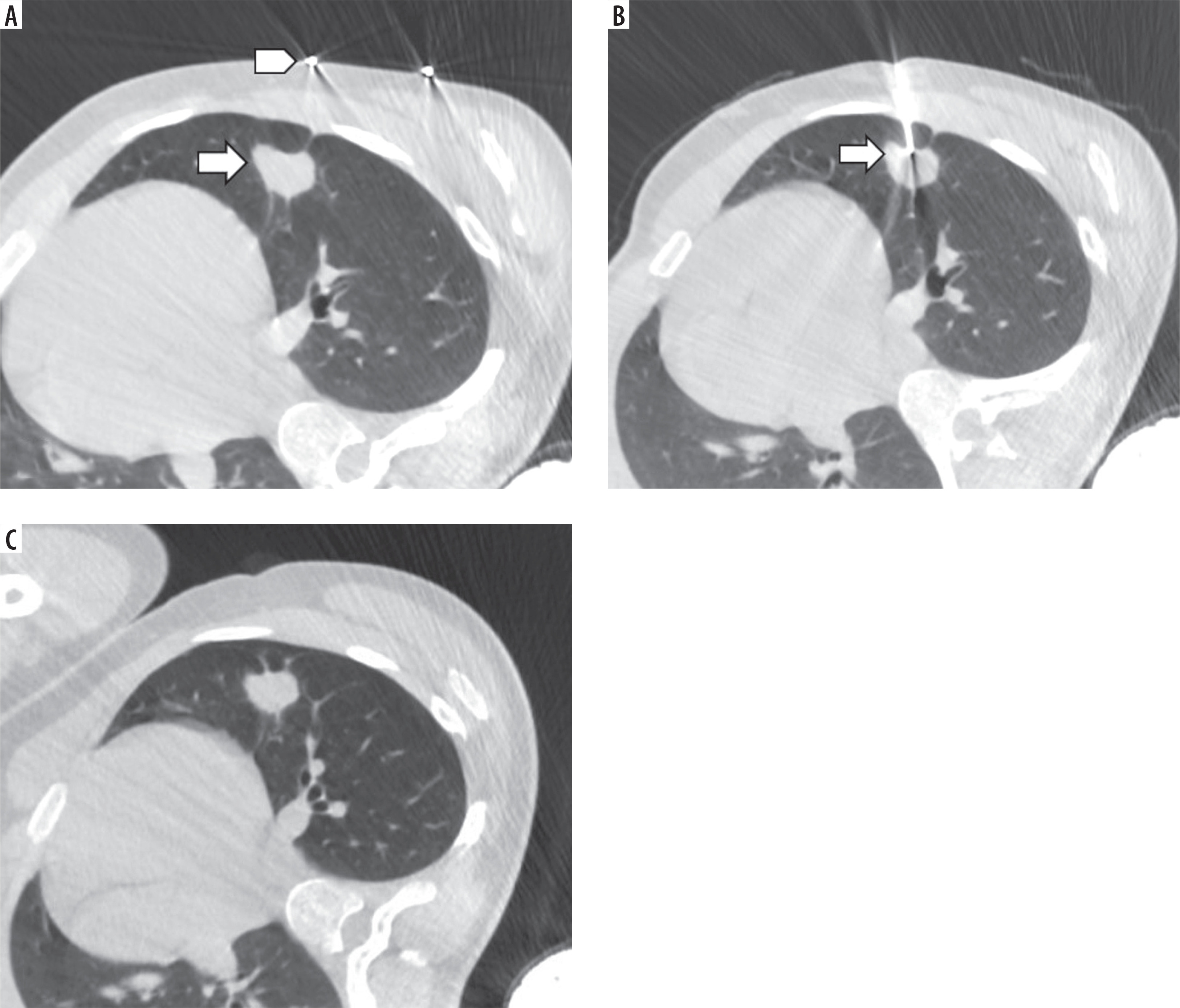
In CT-guided procedures, performed using a 16-slice CT scanner (Optima CT540, GE Healthcare, San Diego, Calif), a radiodense marker was affixed to the patient’s skin, aligned as parallel as possible to the lesion. The lesion levels were scanned in biopsy mode at 120 kVp and 30-150 mAs without contrast injection. The optimal puncture site for the biopsy was identified on the axial CT scan, and the table was adjusted to align this level with the gantry. The mediolateral distance from the radiodense marker to the puncture site was measured, and the skin was marked with a surgical marker at the corresponding distance along the laser light projected onto the designated slice level. After removing the radiodense marker and disinfecting the area, local anesthesia was administered. A 17- or 19-gauge coaxial needle (Geotek Medical, Ankara, Turkey) was inserted to the measured depth to ensure accurate placement within the lesion targeted for biopsy. Intermittent CT imaging was occasionally used to confirm the accuracy of the needle tract. Once the needle was verified to be within the lesion, tissue samples were obtained using an 18- or 20-gauge semi-automatic Tru-Cut biopsy needle. At least two core specimens were obtained, preserved in 10% formalin, and sent for pathological examination. A post-procedure CT scan was performed to detect any potential complications (Figure 2).
Figure 2
An axial computed tomography (CT) scan of a 17-year-old male patient demonstrates a 2.5 cm nodule (arrow) adjacent to the fissure in the left lung, with a radiodense marker placed on the skin parallel to the nodule (arrowhead) (A). The depth between the insertion site and the lesion was measured before advancing the needle into the lesion, and a subsequent CT scan confirmed precise needle placement within the nodule (arrow) (B). Post-procedural CT revealed no complications (C)
During the earlier study period (before October 2016), CT-guided biopsies were performed using a standard-dose protocol (120 kV, 150 mAs) in 4 patients. Following institutional protocol optimization after October 2016, low-dose CT protocols (120 kV, 30 mAs) were adopted and applied in 7 patients. Slice thickness was adjusted according to lesion size: a thinner slice thickness (1.25 mm) was used for lesions smaller than 1.5 cm, whereas a thicker slice thickness (3 mm) was preferred for larger lesions to further reduce radiation exposure.
Patients were placed in a biopsy-side-down lateral decubitus position and monitored for one hour following the procedure. A follow-up chest radiograph was obtained two hours after the procedure. Asymptomatic patients were discharged after an overnight hospitalization.
Definitions and statistical analysis
Technical success was defined as the correct placement of the biopsy needle within the target lesion and successful acquisition of tissue samples.
Statistical analysis was performed using SPSS software, version 26.0 (IBM Corp., Armonk, NY, USA). The Shapiro–Wilk test was used to assess the normality of distribution for continuous variables. Continuous variables were expressed as median and range, while categorical variables were presented as frequencies and percentages. Fisher’s exact test was used to compare categorical variables. Diagnostic performance metrics – including sensitivity, specificity, positive predictive value (PPV), and negative predictive value (NPV) – were calculated using final clinical and/or surgical confirmation as the reference standard. A p-value < 0.05 was considered statistically significant.
Results
A total of 40 pediatric patients (24 males, 16 females; median age: 6 years; age range: 1-17) who underwent image-guided transthoracic biopsy were included in the study. The median lesion size was 42 mm (range: 8-132 mm). US-guided transthoracic biopsy procedures were performed for lesions involving the mediastinum (n = 8), pleura (n = 9), or peripheral lung parenchyma (n = 12). CT guidance was used for biopsies of intraparenchymal lesions located in the central parts of the lung (n = 11). Technical success was achieved in 100% of cases using both US-guided and CT-guided techniques.
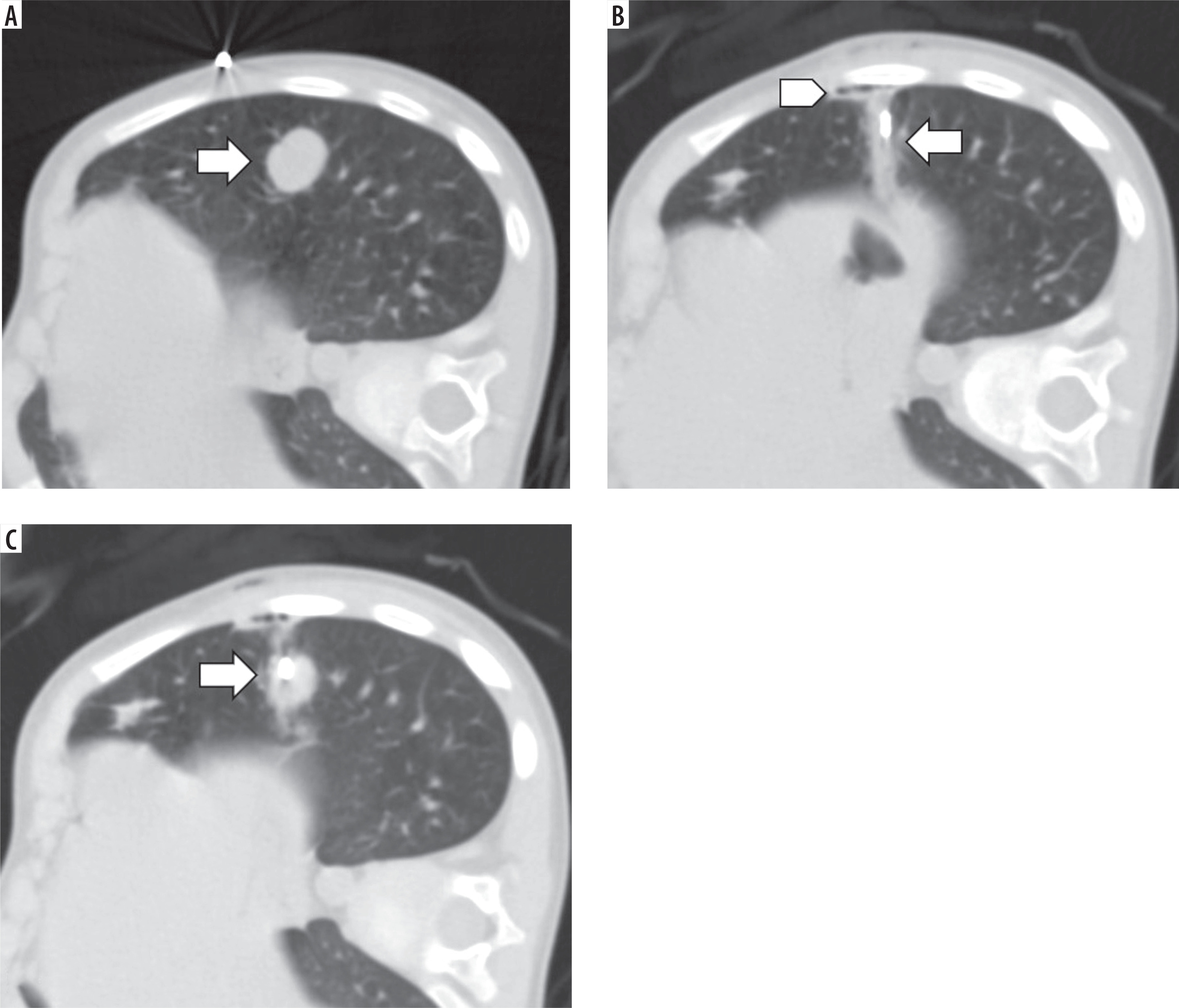
In the CT-guided biopsy group, pneumothorax was observed in two patients (2/11, 18.2%), and focal pulmonary hemorrhage along the needle path was noted in one of these patients (1/11, 9.1%) (Figure 3). Both patients were asymptomatic and managed with nasal oxygen therapy in the biopsy-side-down decubitus position. In the US-guided biopsy group, pneumothorax occurred in two patients (2/29, 6.9%). One of these patients was symptomatic and required chest tube insertion. According to the SIR guidelines [9], this was the only major complication observed in the study, while the other patient was asymptomatic and managed similarly with nasal oxygen therapy and positioning in the biopsy-side-down decubitus position. A retrospective analysis of radiological imaging in 21 patients with biopsy-confirmed malignancy revealed no evidence of tumor seeding along the needle path, with a median interval of 42 months (range: 3-117 months) from biopsy to the most recent imaging. There was no statistically significant difference in the overall complication rates between the CT-guided and US-guided biopsy groups (p = 0.300).
Figure 3
An axial computed tomography scan in the lateral decubitus position of a 2-year-old male patient demonstrates a 17 mm nodule (arrow) in the left lower lobe (A). A scan obtained to confirm needle placement reveals minimal pneumothorax (arrowhead) and pulmonary hemorrhage (arrow) along the coaxial needle path (B). Despite these complications, the patient remained asymptomatic, and after confirming the needle’s placement within the nodule (arrow), tissue samples were successfully obtained (C). The patient was positioned in the biopsy-side-down lateral decubitus position, and nasal oxygen therapy was initiated. After overnight hospitalization, the patient was discharged
Histopathological results are detailed in Table 1. Of the 40 patients, 21 had malignant or borderline lesions, while 18 had benign diagnoses. In two cases, the initial biopsy was non-diagnostic. Following re-biopsy, one patient was diagnosed with plasmablastic lymphoma, and the initial biopsy in this patient was therefore considered false-negative. The other patient did not undergo re-biopsy, as the lesion regressed during this period.
Table 1
Histopathological results obtained from image-guided transthoracic biopsies
Of the 21 patients with malignant or borderline diagnoses based on CNB results, 14 diagnoses were confirmed through surgical resection. The remaining patients, diagnosed with lymphoma, did not undergo surgery but had their diagnoses clinically confirmed based on their response to chemotherapy and other malignancy-related findings. No false-positive cases were identified.
Among the 18 patients with benign diagnoses, 14 were clinically confirmed based on their response to treatment or the absence of lesion growth during a follow-up period of at least one year. One patient’s diagnosis was confirmed through surgical resection, which revealed cystic adenomatoid malformation. Three patients diagnosed with infection or pneumonia were immunocompromised due to common variable immunodeficiency (CVID) and exhibited rapid progressive disease that was incompatible with malignant processes.
The diagnostic accuracy of the procedures was 95% after the initial biopsy procedures and increased to 97.5% following re-biopsy. The sensitivity, specificity, positive predictive value (PPV), and negative predictive value (NPV) of CNB in this series were 95.2%, 100%, 100%, and 94.7%, respectively.
In CT-guided biopsy procedures, dose-length product (DLP) values ranged from 55 to 404 mGy·cm, with a mean value of 181 mGy·cm, corresponding to a mean effective dose of 3.07 mSv.
Discussion
This study demonstrates that image-guided transthoracic CNB is a safe and accurate method for diagnosing thoracic lesions in children. Technical success was achieved in all procedures, and the overall diagnostic accuracy increased to 97.5% following re-biopsy. The complication rate was low, with four cases of pneumothorax (one requiring chest tube insertion) and no statistically significant difference observed between the CT- and US-guided biopsy groups. These findings support the feasibility and reliability of image-guided transthoracic CNB for evaluating mediastinal and pulmonary lesions in the pediatric population.
Data regarding both the incidence and risk factors of pneumothorax following transthoracic biopsy in pediatric patients are limited, and most available evidence originates from studies conducted in adult populations. Therefore, reported pneumothorax rates and associated risk factors derived from adult series may not directly reflect the pediatric population and should be interpreted with caution. Pneumothorax has been reported in 11.6% to 35% of cases following percutaneous transthoracic biopsy, with chest tube placement required in approximately 1% to 14.2% of patients [8,10-14]. Patients with lesions located in the mediastinum or pleura are at minimal risk when aerated lung tissue is not traversed [10]. However, patients with centrally located lung lesions or smaller lesion sizes are at a higher risk [15]. A pneumothorax rate of 52.7% was reported in a study focused on percutaneous biopsy of pulmonary nodules measuring 10 mm or smaller [16].
Technically, the risk of pneumothorax increases with a wider needle insertion angle [11] and an increased number of pleural punctures [17], whereas the use of a smaller coaxial needle helps to reduce it [18]. In our procedures, the insertion angle was planned to be as perpendicular to the pleura as possible, and the biopsy path was carefully designed to avoid traversing fissures, thereby minimizing the number of pleural punctures through the use of the coaxial technique.
The incidence of pneumothorax in this study was 18.2% (2/11) in the CT-guided biopsy group and 6.9% (2/29) in the US-guided biopsy group, consistent with rates reported in the literature [8,10-14]. Although this difference was not statistically significant, the lower pneumothorax rate observed in the US-guided group may partly reflect differences in post-procedural imaging, as CT is more sensitive than chest radiography for detecting minimal pneumothorax. It should be emphasized that CT-guided biopsy was performed in a limited number of patients and was reserved for selected cases in which US guidance was not feasible. Therefore, comparisons between CT-guided and US-guided biopsy techniques should be interpreted with caution and are intended to be descriptive. The chest tube insertion rate was 3.4% (1/29) in the US-guided biopsy group, consistent with previously reported rates in the literature, while no chest tube insertion was required in the CT-guided biopsy group.
The incidence of pulmonary hemorrhage related to transthoracic biopsy has been reported to range between 1% and 27% [10,12,19]. A longer biopsy path and smaller lesion size are associated with an increased risk of bleeding [10,12]. Hemorrhage risk may be higher in patients with emphysema due to reduced tamponade from adjacent tissues [20] and in those with pulmonary arterial hypertension [21]. In the present study, the rate of pulmonary hemorrhage was 9.1% (1/11) in the CT-guided biopsy group. In the US-guided group, no hemorrhage-suggestive infiltration was detected on post-procedural chest radiographs.
Lucidarme et al. [22] reported that CNB provides more accurate diagnostic results compared to FNA while maintaining similar complication rates. Laurent et al. [23] recommended CNB as the primary method for CT-guided lung biopsy, emphasizing its advantages over FNA, including fewer indeterminate samples, improved diagnostic accuracy for both malignant and benign lesions, and a comparable frequency and severity of complications. In this study, CNB was the preferred technique for image-guided percutaneous transthoracic biopsy.
The diagnostic accuracy of transthoracic CNB has been reported to range from 83% to 96% [3,18,22,24,25]. In this study, due to the absence of an on-site cytopathologist, at least two samples were obtained to ensure specimen adequacy. The diagnostic accuracy was 95% after the initial biopsy, increasing to 97.5% following re-biopsy. The sensitivity, specificity, PPV, and NPV were 95.2%, 100%, 100%, and 94.7%, respectively. These findings are consistent with limited studies in pediatric cohorts [26,27] and align with results from large-scale adult cohort studies [3,18,22,24,25].
In this study, US guidance was the preferred modality to avoid exposure to ionizing radiation. The strategy involved using US guidance for mediastinal and pleural lesions. For peripheral intraparenchymal lesions, visibility was initially assessed with US; if the lesion was not visualized, the procedure was then performed under CT guidance. Following institutional protocol optimization in October 2016, CT-guided biopsies were routinely performed using a low-dose protocol in accordance with the ALARA (as low as reasonably achievable) principle. Recent studies have demonstrated no statistically significant difference in diagnostic yield and procedural safety between low-dose CT and standard-dose CT [28,29]. In the present series, procedure-related complications were infrequent in both standard-dose (1/4) and low-dose (1/7) CT-guided biopsy groups. Although the limited sample size precludes formal statistical comparison, these descriptive findings suggest that optimized low-dose CT protocols can preserve procedural safety and diagnostic performance when CT guidance is required. Prioritizing US guidance and applying low-dose CT protocols when necessary provide a diagnostic approach consistent with contemporary principles of pediatric radiation protection, considering children’s increased radiation sensitivity and longer life expectancy.
The limitations of this study include its retrospective design, the lack of comparison with other biopsy techniques, and a relatively small patient cohort, which limits the ability to comprehensively evaluate factors associated with complications or conduct a robust comparison between CT- and US-guided biopsy groups. In addition, this study reflects the experience of a single tertiary referral center, which may limit the generalizability of the results.
In conclusion, image-guided transthoracic biopsies are a safe and reliable technique for the histopathological diagnosis of lung and mediastinal lesions in pediatric patients. This method provides high diagnostic accuracy while maintaining low complication rates.

